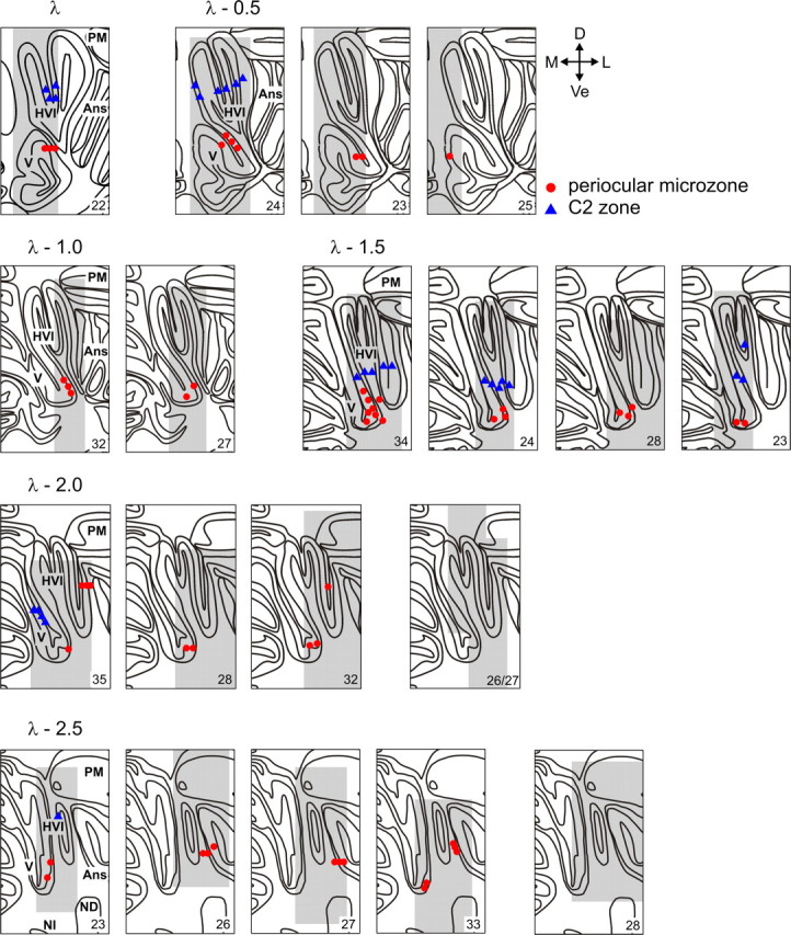
Figure 7.

Summary of the locations of identified climbing fiber zones in rabbit cerebellar cortex across all 11 animals, plotted on a standard series of coronal sections from 0.5 to 2.5 mm caudal to lambda (λ − 0.5 to λ − 2.5). Each panel represents one or more penetrations of the microelectrode array at a single rostrocaudal level in a single animal (sampled area shaded gray). A reference number for the animal is given in the bottom right corner. Each point represents the location on a single electrode track in which the largest-amplitude CFP was recorded (periocular microzones, red; C2 zone, blue). Note the preponderance of periocular microzones located ventrally on the medial wall of lobule HVI down to the base of the primary fissure that separates lobules V and HVI. Identified C2 zones are located dorsal to periocular microzones. Also note a number of illustrated cases at λ − 2.0 mm and λ − 2.5 mm in which the sampled area is shaded but no relevant climbing fiber activity was recorded. Ans, Ansiform lobule; PM, paramedian lobule; ND, dentate nucleus; NI, interposed nucleus; D, dorsal; Ve, ventral; M, medial; L, lateral.
