
Figure 8.
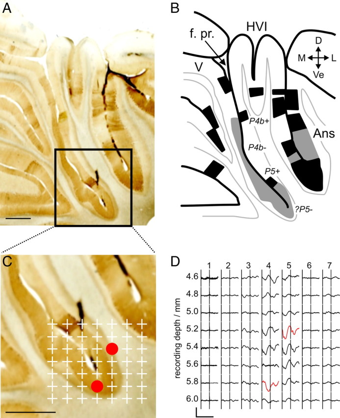
A, Histological section from a mapping experiment (animal #27, λ − 1.0 mm), stained for zebrin immunoreactivity (scale bar, 1 mm). B, Annotated illustration of lobular organization and zebrin staining in A. Black regions are those that are clearly zebrin positive, and regions with weak staining are colored in gray. Zebrin bands are labeled based on the nomenclature of Sanchez et al. (2002). C, Expansion of the region in A indicated by the black box (scale bar, 1 mm). Plus signs mark the location of LFP sampling points from a microelectrode array penetration. Red circles show the location of the largest-amplitude periocular-evoked CFPs. These are illustrated in D; the timing of two periocular stimuli 40 ms apart is indicated by the vertical stimulus artifacts (calibration: 80 ms, 100 μV). Traces in red indicate the largest-amplitude periocular-evoked CFPs in the two electrode tracks in which they were recorded: these were 4.8 and 5.1 mm lateral to the midline. f. pr, Primary fissure; D, dorsal; Ve, ventral; M, medial; L, lateral; Ans, ansiform lobule.