
Figure 9.
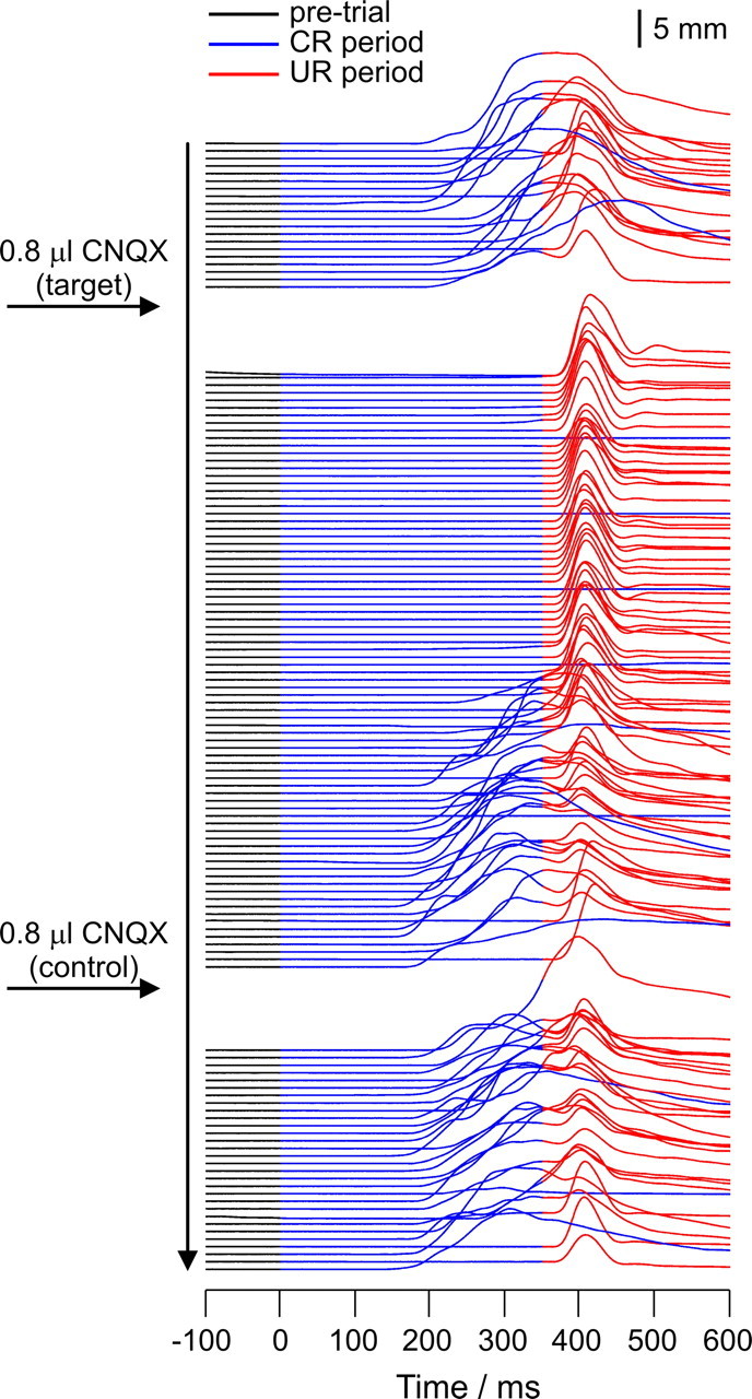
NMRs in a well trained rabbit after infusions of 0.8 μl of 3 mm CNQX into an electrophysiologically identified periocular microzone (target) and into a control region in lobule HVI 4.8 mm dorsally in the same animal (control). CS and US onsets are at times 0 and 350 ms, respectively. Successive trials are drawn in serial order starting from the top. Nictitating membrane positions are drawn as follows: pre-CS, black; CS–US interval, blue; post-US, red. Note that 1 in 10 trials is a CS-alone presentation (entirely blue post-CS). The target infusion transiently abolishes CRs: on CS alone trials, no NMRs are seen at any latency; on paired trials, NMRs within the CS–US interval are abolished but the unconditioned responses remain. The control infusion has no effect. Summary behavioral data for these infusions are presented in Figure 10A.