
Figure 4.
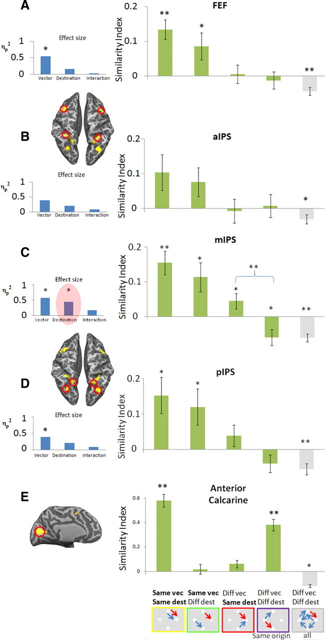
Similarities of spatial patterns of responses in FEF (A), three different subregions of the IPS (B–D), and the anterior calcarine (E). The figure shows mean similarity indexes for different comparisons of delay-period response patterns in the studied ROIs (depicted by the red circles in the partially inflated brain; dorsal view, A–D; medial view, E). The icons in the bottom, below each bar, represent the paired conditions whose average index is depicted above. These include the following (from left to right): identical conditions; same vector; same destination, same origin; and completely different conditions. Blue and red arrows are specific examples of such cases from the separate (odd and even) datasets. The graphs in the insets (light blue bars) show the magnitude of the specific effects (saccade vector, saccade destination, and interaction), assessed by ANOVA (partial η2) on the similarity indexes indicated by the green bars in each graph. Error bars denote SEM across subjects (n = 12). *p < 0.05; **p < 0.005. Note the significant effect of saccade destination in the middle IPS marked in red.