
Figure 3.
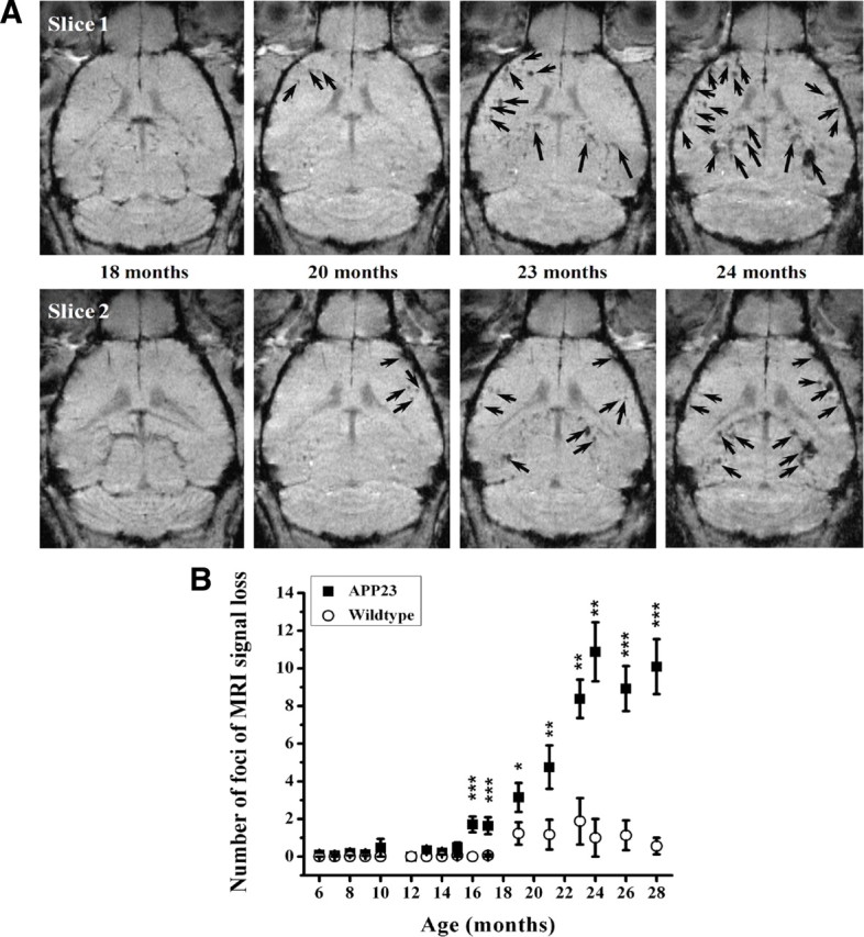
Age-related effects in male APP23 mice. A, Foci of attenuated signal are shown in two slices extracted from 3D MRI datasets acquired at different ages from a representative animal. SPIO was administered 24 h before each image acquisition. B, Age-dependent increase in the number of sites presenting signal attenuation in the brain cortex of male APP23 mice and age-matched littermate controls as determined from 3D MRI datasets. Data are given as mean ± SEM (n = 8–16 mice at each time point). The levels of significance *0.01 < p < 0.05, **0.001 < p < 0.01, and ***0.0001 < p < 0.001 refer to Mann–Whitney statistical tests performed between APP23 and wild-type mice at each specified age. The contrast agent, SPIO, was administered 24 h before each image acquisition.