
Figure 1.
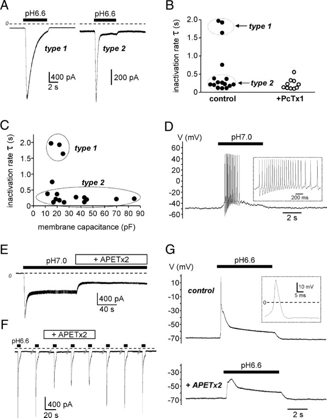
Electrophysiological characterization of ASIC channels in DRG neurons innervating the rat hindpaw muscles. A, Typical ASIC currents recorded in voltage-clamp (holding potential −80 mV) from retrogradely labeled sensory neurons innervating hindpaw muscles. Current was activated by a rapid switch of the external pH from 7.4 to 6.6, as indicated by the black bars above the current traces. The dashed line represents the zero current level. According to their inactivation rates (τ), two main types of ASIC current can be distinguished (type 1 and type 2). B, Analysis of the inactivation rate of native pH 6.6-induced ASIC currents in control condition and in the presence of the PcTx1 toxin. The slow-inactivating type 1 current is blocked by the toxin. C, Functional distribution of type 1 and type 2 ASIC currents as a function of the neuron membrane capacitance. D, Current-clamp experiment performed on a neuron exhibiting a type 2 ASIC current. A slight acidification of the external medium to pH 7.0 depolarizes the membrane sufficiently to trigger firing. Action potentials are magnified in the inset. E, Effect of APETx2 on recombinant sustained ASIC3 current recorded at −80 mV from F11-transfected cells. Current was activated by external pH changes from pH 7.4 to 7.0, as indicated by the black bar. The APETx2 toxin was applied externally at 1 μm, as indicated. F, Effect of APETx2 (5 μm) on native pH 6.6-evoked ASIC current recorded at −80 mV from retrogradely labeled sensory neurons innervating hindpaw muscles. The fast component of the current (type 2) is largely inhibited by the toxin. G, Current-clamp experiment performed on the same neuron as in F. The depolarization induced by the pH 6.6-evoked ASIC current was sufficient to trigger an action potential (magnified in the inset). The presence of APETx2 inhibits the pH 6.6-evoked depolarization and the action potential threshold is not reached.