
Figure 3.
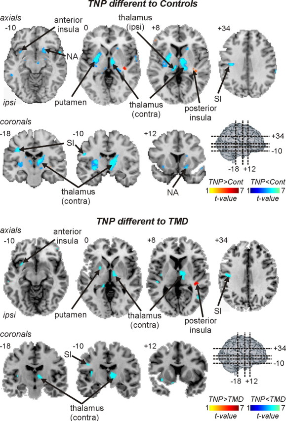
Regional gray matter volume decreases (cool color scale) and increases (hot color scale) in TNP patients compared with controls and compared with TMD patients. Gray matter volume changes are overlaid onto axial and coronal slices of an individual subject's T1-weighted anatomical image set. Slice locations are indicated on the 3-D brain in the top left and by the Montreal Neurological Institute space at the top left of each slice. Note that, compared with both control and TMD subjects, TNP patients have lower gray matter volumes in the ipsilateral (ipsi) anterior insula, putamen and primary somatosensory cortex (SI), and the contralateral (contra) thalamus. Furthermore, compared with control subjects, TNP patients also had decreased gray matter volume in the ipsilateral thalamus and the contralateral nucleus accumbens (NA).