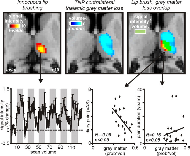
Figure 4.
Gray matter volume decreases in the contralateral thalamus of TNP patients and contralateral (contra) thalamic activity increases during innocuous brushing of the lower lip overlaid onto axial slices of an individual subject's T1-weighted anatomical image set. Light blue indicates gray matter volume decrease, red indicates activity increases during lip brushing, and yellow indicates where both gray matter volume decreased and activity (signal intensity) increased. Slice location in Montreal Neurological Institute space is indicated on the bottom right of the left slice. Bottom left shows mean ± SEM percentage change in signal intensity plotted against time for the contralateral thalamus. The gray bars indicate the periods during which the lower lip was lightly brushed with a plastic brush. Bottom middle and right show plots of gray matter volume decrease (prob * vol) in the area of overlap (i.e., yellow shading, the ventroposterior thalamus) against ongoing diary pain, as indicated on a continuous visual-analog scale (VAS) (bottom middle) and against pain duration (bottom right). Note the significant negative correlation between gray matter volume and pain, i.e., the greater the pain, the greater the reduction in gray matter volume.

