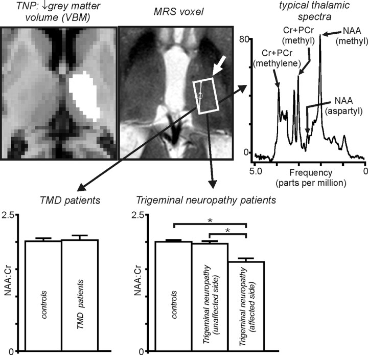
Figure 6.
Top, Thalamic region in which gray matter volume decreased in TNP patients and region from which H1-spectra were collected. To the right is a typical H1-spectra. PCr, Phosphocreatine. Bottom, Left, NAA/Cr within the ventroposterior thalamus in controls, TMD, and trigeminal neuropathy patients on the side ipsilateral (unaffected side) and contralateral (affected side) to ongoing pain. Note that, although there is no significant change in NAA/Cr in TMD patients, NAA/Cr is significantly reduced in the thalamus on the side contralateral to the ongoing pain in trigeminal neuropathy patients. *p < 0.05.

