
Figure 1.
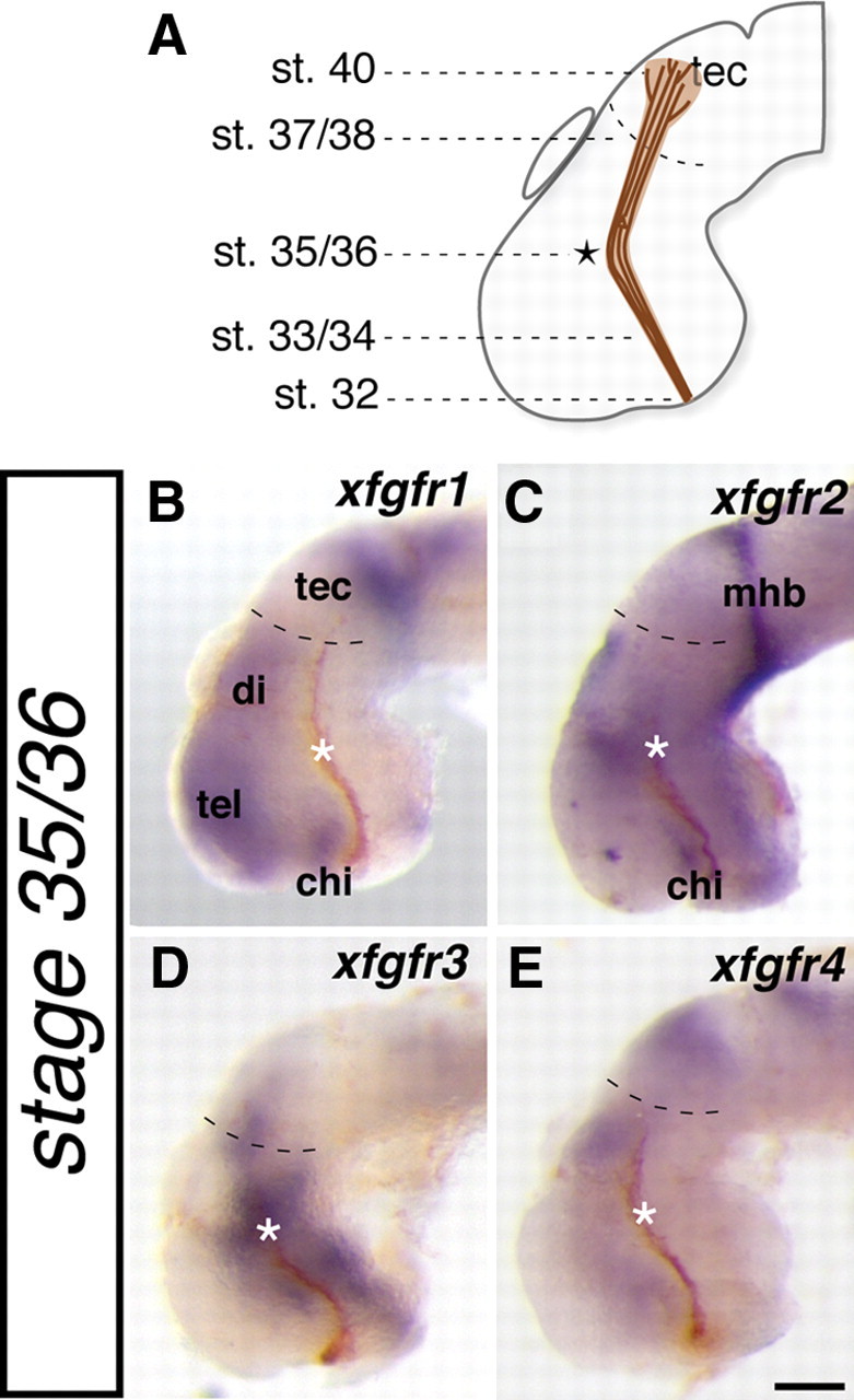
fgfrs are expressed adjacent to the developing optic tract. A, Schematic illustrating where along the optic tract the first RGC axons have reached at each stage of embryonic development. B–E, Lateral whole-mount views of stage 35/36 X. laevis brains in which expression of four fgfrs (xfgfr1–xgfgr4) was determined by in situ hybridization (blue staining). HRP followed by a DAB reaction was used to anterogradely label RGC axons from the contralateral eye (brown fibers). Asterisks indicate the location where RGC axons make a caudal turn in the mid-diencephalon, and dashed lines in B–E delineate the approximate rostral boundary of the optic tectum. chi, Optic chiasm; di, diencephalon; mhb, midbrain–hindbrain border; tec, optic tectum; tel, telencephalon. Scale bar, 100 μm.