Figure 4.

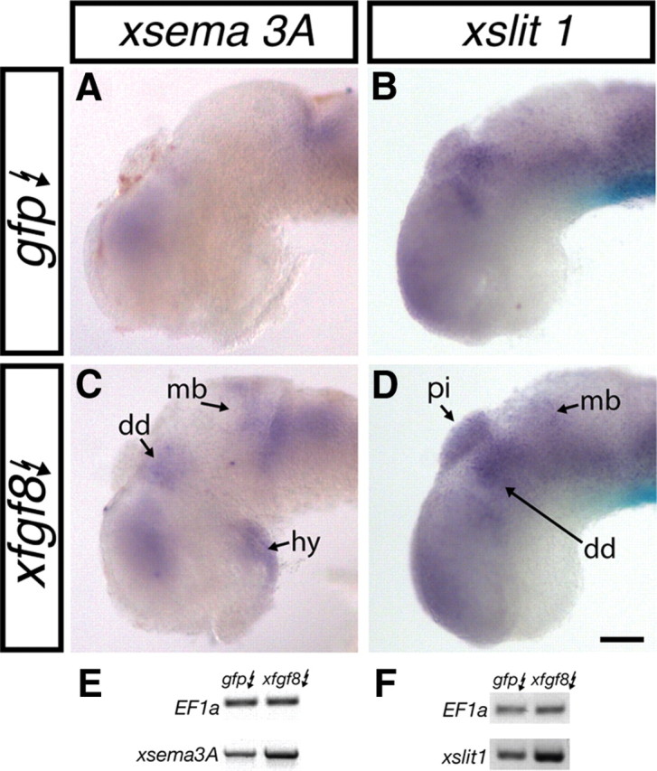
FGF signaling is sufficient to induce xsema3A and xslit1 expression. A–D, Stage 28 embryos were electroporated with pCS2–gfp (gfp) (A, B) or pCS2–gfp and pCS2–xfgf8 (xfgf8; C, D). Twenty-four hours later, embryos were processed for xsema3A (A, C) or xslit1 (B, D) expression by in situ hybridization. Regions of expanded or premature expression are indicated with arrows. E, F, Representative images of the change in xsema3A (E) and xslit1 (F) mRNA levels after gfp and xfgf8 electroporation as assessed by RT-PCR. dd, Dorsal diencephalon; hy, hypothalamus; mb, midbrain; pi, pineal gland. Scale bar, 100 μm.
