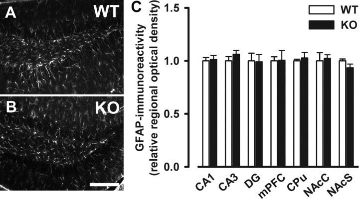
Figure 8.
No indication of reactive gliosis in Nogo-A−/− mice. A, B, Representative images of immunostaining with the astrocyte marker GFAP in the dentate gyrus of Nogo-A+/+ and Nogo-A−/− mice. C, Quantitative analysis of the GFAP staining. No difference between Nogo-A−/− and Nogo-A+/+ mice was detected in the relative optical density of GFAP. All values are mean ± SEM. Scale bar, 200 μm. DG, Dentate gyrus; mPFC, medial prefrontal cortex; CPu, dorsal striatum; NAcC, nucleus accumbens core; NAcS, nucleus accumbens shell; KO, Nogo-A−/− (n = 4–6); WT, Nogo-A+/+ (n = 4–6).

