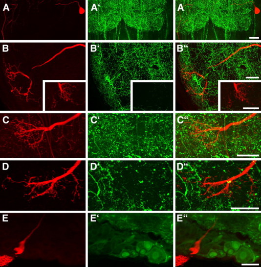
Figure 5.

Confocal images of 130 μm sections showing parts of Neurobiotin-stained descending SOG neurons (red) combined with immunostaining using antisera against FMRFamide, serotonin, Lom-TK II, AST-A, or GABA (green). The Neurobiotin-stained somata and ramifications are shown in A–E, the immunostainings are shown in A′–E′, and the combined illustrations are shown in A‴–E‴. None of the tested neuroactive substance is localized in the stained neurons. A, Soma and part of the primary neurite in the SOG. FMRFamide immunostaining is not present in the soma or axon of the neuron. B, Dendritic ramifications in the SOG and presynaptic terminals from the Pro-TG (inset). The labeled neuron does not show serotonin immunostaining. C, D, In the Pro-TG, presynaptic terminals with bleb-like endings do not show immunostaining for Lom-TK II (C) or AST-A (D). E, The soma of the SOG neuron does not exhibit GABA immunostaining. Scale bars, 40 μm.
