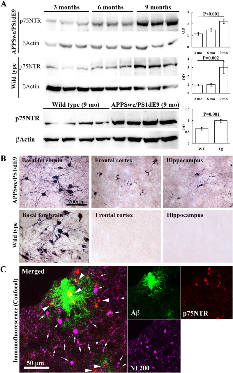
Figure 1.

Expression of p75NTR in the brain. p75NTR expression in the brain was measured by Western blot and immunohistochemistry (n = 10 in each group). A, Brain homogenates of APPSwe/PS1dE9 mice and their wild-type littermates at 3, 6, and 9 months of age were subjected to Western blot analysis probed with rabbit anti-p75NTR polyclonal antibody (G3231) and monoclonal antibody to β-actin. B, Sections of basal forebrain, frontal lobe, and hippocampus from 9-month-old APPSwe/PS1dE9 mice and their wild-type littermates were stained using free-floating immunohistochemistry for p75NTR with rabbit anti-p75NTR polyclonal antibody (Ab9650). C, Representative confocal images for colocalization of p75NTR-positive fibers and fibrillar plaques in brain of 9-month-old APPSwe/PS1dE9 mice, with Ab 9650 for p75NTR (arrowheads), N52 for neurofilament 200 (NF200, arrows), and thioflavine S for fibrillar plaque.
