Figure 6.

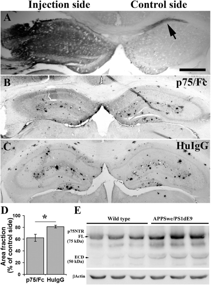
Hippocampus injection of p75/Fc reduces local Aβ plaques. p75/Fc (3 μg in 3 μl) or HuIgG (6 μg in 3 μl, the equivalent molar to p75/Fc) were injected into the left hippocampus of 9-month-old APPSwe/PS1dE9 mice (n = 4 in each group). One week after injection, Aβ plaques in hippocampus were stained using biotin-conjugated 6E10 antibody and quantified. The area fraction of Aβ plaque in hippocampus of the injection side was normalized with the control side. A, Distribution and diffusion of p75/Fc 24 h after injection in the left hippocampus. Sections were stained with antibody to Fc of human IgG. B, C, Representative images of hippocampus Aβ plaque staining 7 d after injection of p75/Fc (B) or HuIgG (C) into the hippocampus of 9-month-old APPSwe/PS1 mice. D, Comparison of Aβ plaque burden in hippocampus between p75/Fc and HuIgG injection groups. E, Expression of p75NTR extracellular domain (ECD) in the brain of wild-type and APPSwe/PS1dE9 mice at age of 9 months. To see the diffusion of injected p75/Fc in the hippocampus, three mice were killed 24 h after injection and brain sections were stained against Fc fragment of human IgG. Extensive diffusion of the protein was observed in the injected hippocampus, whereas little protein diffused into the contralateral hippocampus (arrow). * denotes p < 0.05.
