
Figure 2.
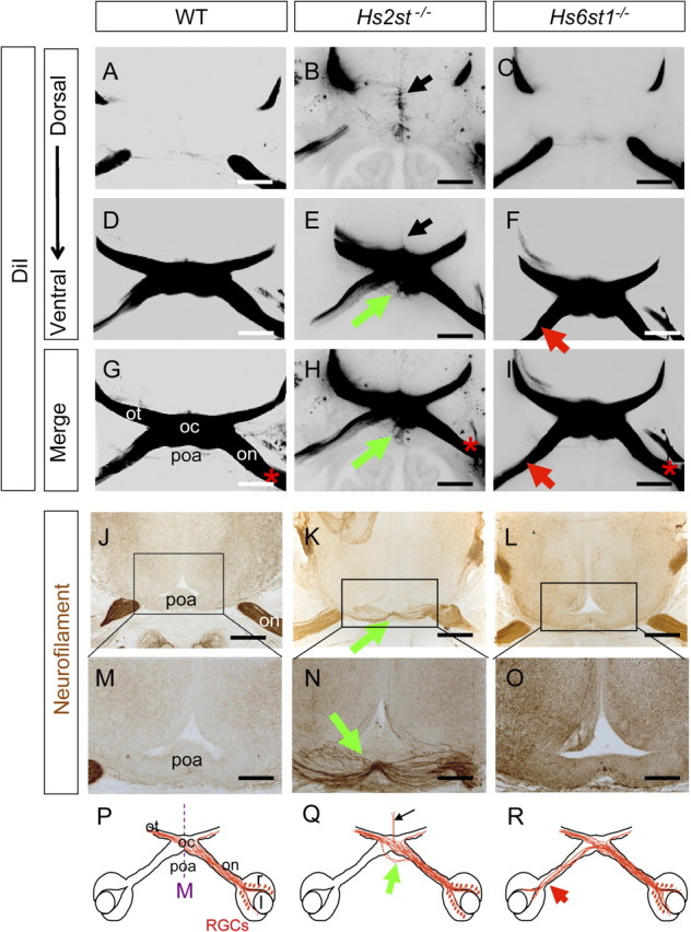
Ectopic retinal ganglion cell axon growth through the preoptic area in Hs2st−/− embryos. A–F, DiI injection into the retina labeling RGC axons at the optic chiasm in E15.5 embryos. DiI images are presented as grayscale negatives for clarity. B, E, Hs2st−/− embryos RGC axons escape the normal chiasm region and grow ectopically across the preoptic area (E, green arrow) as well as dorsally up the midline (B, black arrow). This ectopic axon growth does not occur in wild-type (A, D) or Hs6st1−/− (C, F) embryos. G–I, Merges of A and D (G), B and E (H), and C and F (I), with * marking side of DiI application. J–O, RGC axons stained brown using neurofilament immunohistochemistry. J–O, E15.5. Wild-type embryo (J, M), Hs2st−/− embryo (K, N), Hs6st1−/− embryo (L, O). An ectopic branch of the optic nerve enters the preoptic area in Hs2st−/− embryos (K, N, green arrows) which is absent in the other genotypes. P–R, Diagrams summarizing RGC axon navigation at the chiasm of wild-type, Hs2st−/−, and Hs6st1−/− embryos; green arrows, preoptic area tract; black arrows, midline wandering; red arrow (R), increased inter-retinal innervation previously described (Pratt et al., 2006). on, Optic nerve; oc, optic chiasm; ot, optic tract; r, retina; l, lens; poa, preoptic area; M, midline. All sections horizontal with caudal at top. Scale bars: A–L, 500 μm; M–O, 250 μm.