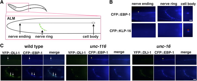
Figure 2.
Localization of DLI-1 in ALM touch neurons. A, Schematic image of ALM touch neuron. Presynapses are indicated by green. B, Localization of CFP::EBP-1 and CFP::KLP-16 in ALM neurons. The yellow arrowhead indicates the location of CFP::EBP-1 at the nerve ending of ALM neuron. The red arrowhead indicates the accumulation of CFP::KLP-16 in the cell body. C, Localization of YFP::DLI-1 and CFP::EBP-1 in ALM neurons in wild-type, unc-116(e2281), and unc-16(e109) mutant worms. The top and bottom panels show the processes and nerve rings of ALM neurons, respectively. The arrows indicate the positions of colocalized YFP::DLI-1 and CFP::EBP-1 at the ends of ALM neuron of processes and nerve rings. The yellow arrowheads indicate the locations at the nerve endings. The merged images are shown in right panels. Scale bars, 10 μm.

