Figure 4.
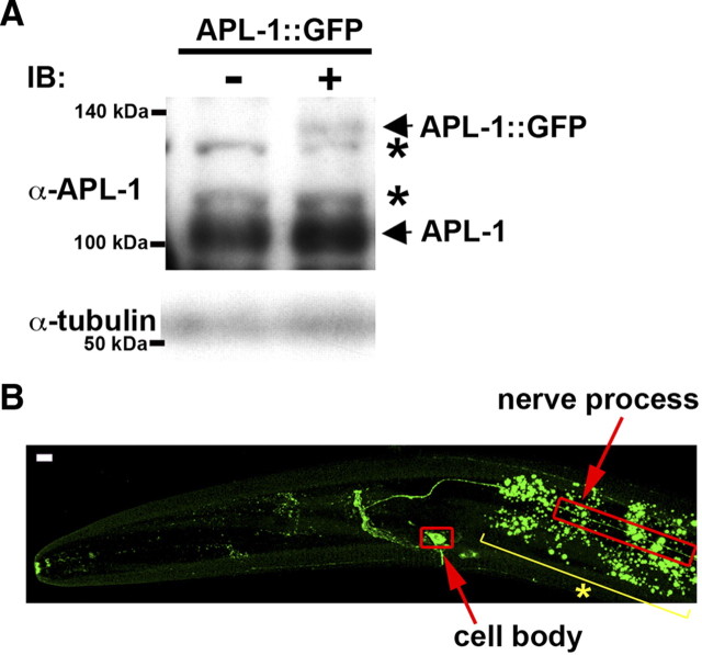
Expression and localization of APL-1::GFP. A, Immunoblotting of APL-1 proteins. Whole extracts were prepared from worms at mixed stages. The top panels show immunoblotting with anti-APL-1 antibody. Positions of APL-1::GFP and endogenous APL-1 are indicated by arrows. The asterisks indicate nonspecific bands. The bottom panels show immunoblotting with anti-tubulin antibody as a loading control. B, Image of fluorescent APL-1::GFP protein in the heads of L4 larva. The red open boxes indicate the cell body and nerve processes of the sublateral motor neurons. The yellow line with an asterisk indicates gut fluorescent granules. Images are from a confocal Z series projected onto a single plane. Scale bar, 5 μm.

