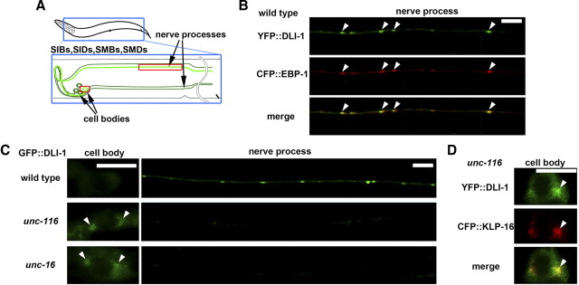
Figure 5.
Localization of DLI-1 in sublateral motor neurons. A, Schematic image of sublateral motor neurons. SIB, SID, SMB, and SMD neurons are indicated by green lines. B, Localization of YFP::DLI-1 and CFP::EBP-1 in the nerve processes of sublateral neurons. The merged image is also indicated. The arrowheads indicate the merged sites. Scale bar, 5 μm. C, Localization of GFP::DLI-1 in the sublateral neurons. The left and right panels show cell bodies and nerve processes, respectively. The arrowheads indicate the accumulated positions of GFP::DLI-1 in cell bodies. Scale bar, 5 μm. D, Localization of YFP::DLI-1 and CFP::KLP-16 in the cell bodies of sublateral neurons in unc-116(e2281) mutants. The arrowheads indicate the merged sites.

