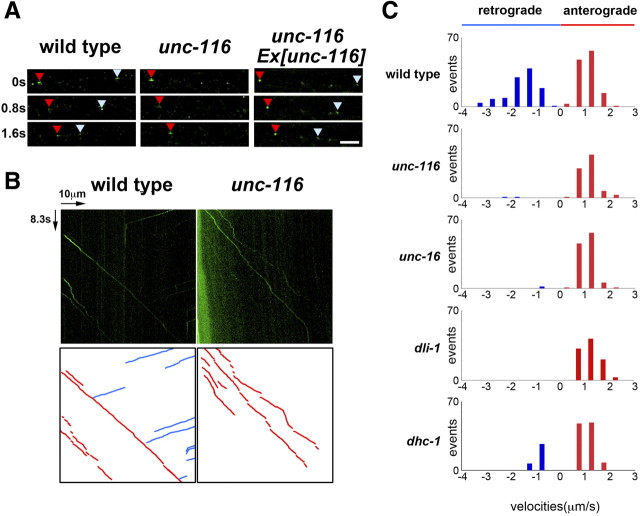
Figure 8.
Movement of APL-1 in sublateral motor neurons. A, Time-lapse images of APL-1::GFP fluorescence along the sublateral neurons of worms. Each photograph was taken at 0.8 s intervals. The red and pale blue arrowheads indicate APL-1::GFP vesicles moving toward neurite endings (anterograde) and cell bodies (retrograde), respectively. Scale bar, 2.5 μm. B, Kymographs for APL-1::GFP movements along the lateral processes. The horizontal arrow represents 10 μm. The vertical arrow represents 8.3 s. Schematic diagrams are shown below. The red and blue lines represent anterograde and retrograde movements, respectively. C, Velocity profiles of moving vesicles containing APL-1::GFP in wild-type, unc-116(e2281), unc-16(e109), dli-1(ku266), and dhc-1(or195ts) worms. Moving structures were tracked, and the velocities were calculated as described in Materials and Methods.

