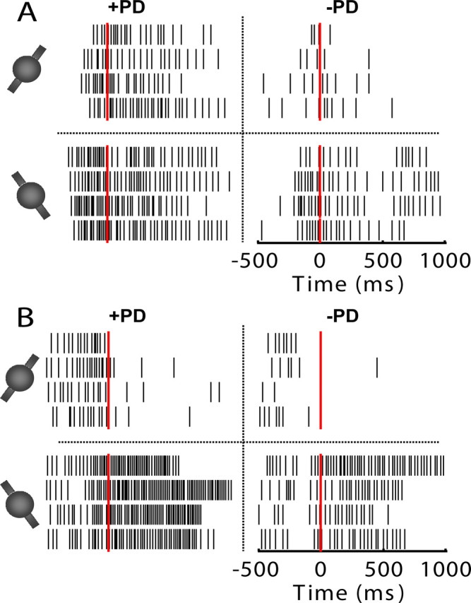
Figure 2.

Spike rasters of two representative motor cortical neurons tuned to both translation and rotation from two separate monkeys (A, B). All rasters are aligned to movement onset time (time 0, red line). Thin black vertical lines indicate occurrences of spikes from 0.5 s before movement onset to 1 s after movement onset. Rasters are divided into four groups by the dotted lines. Those four groups correspond to four targets, i.e., four different combinations of translation and rotation. The top and bottom rows correspond to movements with CW and CCW rotations, respectively. The left and right columns correspond to movements along the preferred translational direction (+PD) and movements opposite to the preferred translational direction (−PD), respectively. Difference in spike rate between the left and right columns indicates translational tuning, while difference in spike rate between the top and bottom rows indicates rotational tuning.
