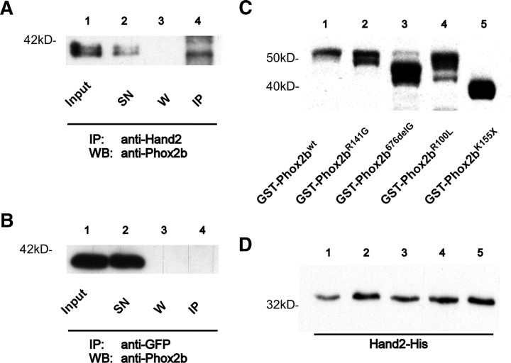
Figure 5.
Protein–protein interaction between Phox2b and Hand2. A, B, Coimmunoprecipitation of Phox2b and Hand2 from SY5Y neuroblastoma cells. Western blots with antibodies specific for Phox2b after immunoprecipitation with anti-Hand2 (A) or anti-GFP (B) antibodies. Input, Input control; SN, supernatant after agarose bead and Hand2 (A) or GFP (B) antibody incubation; W, supernatant after washing immunopellet with lysis buffer; IP, immunopellet. Anti-GFP antibody (B) and agarose beads alone (data not shown) served as negative controls. Phox2b bands with different apparent molecular weights most likely represent differentially phosphorylated forms (Adachi and Lewis, 2002). C, D, Analysis of Phox2b–Hand2 interaction by pulldown experiments. Bacterial lysates of GST-Phox2b fusion proteins were first incubated with GST-beads and subsequently with Hand2 bacterial lysate. C, Western blot analysis of GST-Phox2b fusion proteins that were used for the pulldown of Hand2 visualized by GST-HRP-coupled antibodies. D, Hand2-His was detected by antibodies against the His-tag and is present in every lane after GST-pulldown with GST-Phox2b fusion proteins. GST-beads alone and GST-Protein served as negative controls whereas Hand2 protein lysate served as positive control for specific protein–protein interaction (data not shown).

