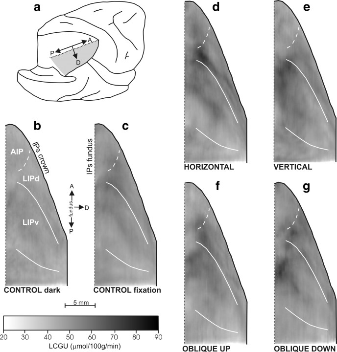
Figure 3.
Two-dimensional reconstructions of the distribution of metabolic activity in area LIP. Quantitative maps of the spatiointensive distribution of LCGU values (in micromoles · 100 g−1 · minute−1) in the lateral bank of the IPs. a, Posterolateral view of the partially dissected right hemisphere of a monkey brain. The IPs was unfolded after the inferior parietal lobule was cut away below the posterior (lateral) crown of the IPs, and the occipital lobe was cut away at the fundus of parietoccipital and lunate sulci. The shaded area represents the reconstructed lateral (lower) bank of the IPs. b, Two-dimensional map of activity in the cortex of the reconstructed lateral bank of the IPs, averaged from the four hemispheres of the two control monkeys in the dark (Cd). The solid black line on the right represents the crown of IPs; the vertical dotted black line on the left depicts its fundus. The solid white lines correspond to the cytoarchitectonically identified borders of area LIPv. The dashed white line corresponds to the functionally identified border between LIPd and AIP (Evangeliou et al., 2009). c, Two-dimensional map of the lateral bank of IPs, averaged from the four hemispheres of the two control fixating monkeys (Cf). d, Averaged map from three hemispheres of monkeys executing contraversive horizontal saccades of 5, 10, 15, and 30° amplitude. e, Averaged map from two hemispheres of the monkey executing vertical saccades of 5, 10, 15, and 30° amplitude up and down. f, Averaged map from four hemispheres of monkeys executing contraversive saccades of 10, 20, and 30° amplitude from straight ahead in an oblique direction 135° up. g, Averaged map from three hemispheres of monkeys executing contraversive saccades of 10 and 20° amplitude from straight ahead in an oblique direction 315° down. Gray-scale bar indicates LCGU values in micromoles · 100 g−1 · minute−1. A, Anterior; AIP, anterior intraparietal area; D, dorsal; P, posterior.

