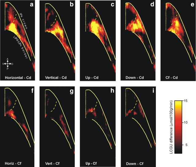
Figure 9.
Effects in LIPd, expressed as LCGU differences relative to Cd and to Cf monkeys. a, Quantitative two-dimensional map of activity in the LIPd of monkeys executing horizontal saccades, averaged over the three hemispheres of monkeys H1 and H2 shown in Figure 4, after subtracting the average of the four hemispheres of the Cd monkeys. b, Reconstructed two-dimensional map of activity in the LIPd averaged over both hemispheres of a monkey executing vertical saccades, minus the Cd control map. c, Two-dimensional map of the average activity in the LIPd of four monkeys executing oblique upward saccades averaged over all four contralateral hemispheres, minus the Cd control map. d, Reconstructed two-dimensional map of the average activity in the LIPd of three monkeys executing oblique downward saccades, averaged over all three contralateral hemispheres, minus the Cd control map. e, Reconstructed two-dimensional map of the activity in the LIPd of Cf monkeys averaged over four hemispheres, minus the Cd control map. f, Quantitative two-dimensional map of activity in the LIPd of monkeys executing horizontal saccades, after subtracting the average of the four hemispheres of the Cf monkeys. g, Reconstructed two-dimensional map of activity in the LIPd averaged over both hemispheres of a monkey executing vertical saccades, minus the Cf map. h, Two-dimensional map of the average activity in the LIPd of four monkeys executing oblique upward saccades, minus the Cf map. i, Reconstructed two-dimensional map of the average activity in the LIPd of three monkeys executing oblique downward saccades, minus the Cf map. The color bar indicates LCGU differences from the control (Cd or Cf) in micromoles · 100 g−1 · minute−1. The location and orientation of LIPd relative to other portions of the lateral bank of the IPs is shown in Figure 4. The vertical straight yellow line on the left is the fundus of the IPs. The upper solid yellow line on the right is the crown of the IPs, the lower solid yellow line is the cytoarchitectonically identified border between LIPv and LIPd, and the dashed yellow line is the functionally identified border between LIPd and AIP (Evangeliou et al., 2009).

