
Figure 4.
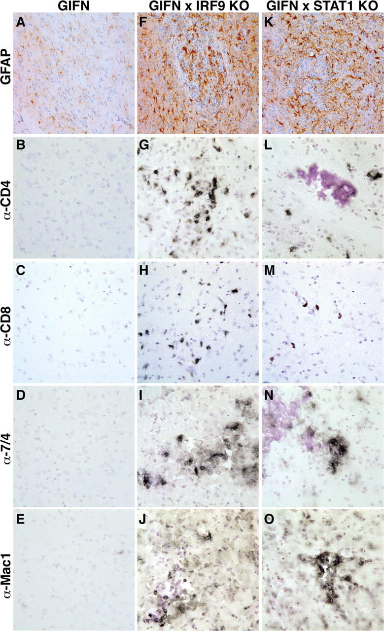
Leukocytes in the CNS of GIFNxSTAT1 KO and GIFNxIRF9 KO mice consisted of T cells, macrophages/monocytes, and neutrophils. A–E, The medial–ventral forebrain of 12-week-old GIFN mice showed no reactive astrocytosis (A) or infiltrating CD4+ (B) or CD8+ T cells (C), neutrophils (D), or macrophages (E). F–I, Pronounced reactive astrocytosis (F) was present in the medial–ventral forebrain of GIFNxIRF9 KO mice, and leukocyte infiltrates consisted of CD4+ (G) and CD8+ (H) T cells and some neutrophils (I). J, Pronounced microglia/macrophage accumulation was evident. K–O, In GIFNxSTAT1 KO mice reactive astrocytosis (K) was similar to GIFNxIRF9 KO mice. Significant numbers of infiltrating CD4+ (L) and CD8+ (M) T cells, neutrophils (N), and microglia/macrophage accumulation (O) were observed in the medial–ventral forebrain of GIFNxSTAT1 KO mice. Original magnification 400×.