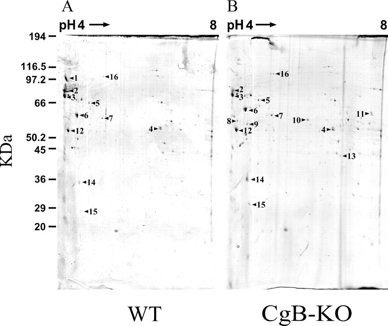
Figure 4.
Two-dimensional gel electrophoresis of chromaffin secretory vesicles from WT and CgB-KO mice. Fractions 3 to 5 from Optiprep gradients were used to perform the 2D SDS-PAGE. Panels show original gels stained with colloidal Coomassie blue. Proteins were identified by MALDI-TOF (see Table 3).

