
Figure 2.
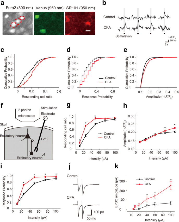
Intracortical remodeling between the L2/3 and L4 in the S1 contributes to the hypersensitivity of L2/3 excitatory neurons to sensory stimulation under chronic pain conditions. a, Visualization of L2/3 excitatory neurons (red dotted circles in the fura-2 image). Cells expressing Venus (inhibitory neurons, green) and cells stained with SR101 (astrocytes, red) were excluded from the analysis. Scale bar, 12 μm. b, Typical traces of sensory-evoked Ca2+ transients of L2/3 excitatory neurons in a control (upper traces) and CFA-injected (lower traces) mouse. c–e, Cumulative probability histograms of the responding cell ratio (c), response probability per cell (d), and amplitude of Ca2+ transients (e). In each analysis, a significant difference between control and CFA-injected mice was observed (Kolmogorov–Smirnov test, p < 0.05). f, Schematic drawing of in vivo Ca2+ imaging of L2/3 excitatory neurons in response to L4 electrical stimulation. g–i, The relationship between stimulus intensity and responding cell ratio (g), amplitude of Ca2+ transients (h), and response probability per cell (i). Excitability of L2/3 excitatory neurons of S1 evoked by electrical stimulation of the L4 region increased in the CFA-injected mice. j, Averaged traces of EPSCs evoked by paired-pulse stimulations of 60 μA in control (top) and CFA-injected (bottom) mice. k, Stimulus intensity–response curves from control (7 cells, filled squares) and CFA-injected (8 cells, filled circles) mice. *p < 0.05, significantly different from the control group by ANOVA followed by Bonferroni test. Error bars represent ±SEM.