
Figure 8.
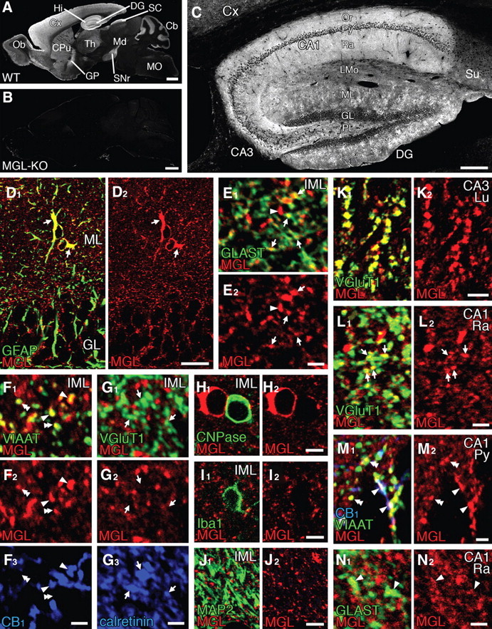
MGL is expressed in astrocytes and some inhibitory terminals in the dentate gyrus and hippocampus. A–C, Immunofluorescence for MGL in parasagittal brain sections of wild-type (WT; A, C) and MGL-knock-out (KO; B) mice. C, An enlarged view of the hippocampus formation. D, E, Double immunofluorescence for MGL (red) and astrocytic marker GFAP (D, green) or GLAST (E, green) in the inner molecular layer (IML) of the dentate gyrus. Note the overlap of MGL with GFAP in perikarya and shaft processes of astrocytes (D, arrows) and with GLAST in their peripheral processes (E, arrows). The arrowhead in E indicates intense MGL in GLAST-immunonegative elements (putative inhibitory terminals). F, Triple immunofluorescence for MGL (red), VIAAT (F1, green), and CB1 (F3, blue) in the IML. Note that intense MGL is found in many VIAAT-positive inhibitory terminals, which include both CB1-positive (arrowheads) and CB1-negative (double arrowheads) terminals. G, Triple immunofluorescence for MGL (red), VGluT1 (G1, green), and calretinin (G3, blue) in the IML. Note that there is no MGL immunoreactivity in MC terminals colabeled for VGluT1 and calretinin (arrows). H–J, Double immunofluorescence for MGL (red) with CNPase (H, green), Iba1 (I, green), or MAP2 (J, green). Note that there is no MGL labeling in CNPase-positive oligodendrocytes, Iba1-positive microglia, or MAP2-positive neuronal dendrites. K, L, Double immunofluorescence for MGL (red) and VGluT1 (green) in the hippocampal CA3 (K) and CA1 (L). M, Triple immunofluorescence for MGL (red), VIAAT (green), and CB1 (blue) in the hippocampal CA1. N, Double immunofluorescence for MGL and GLAST in the hippocampal CA1. In the CA3, intense immunoreactivity is detected in VGluT1-labeled mossy fiber terminals (K). In the CA1, MGL immunoreactivity is detected in VGluT1-labeled excitatory terminals of Schaffer collaterals (L, arrows), VIAAT-labeled inhibitory terminals including both CB1-positive (M, arrowheads) and CB1-negative (M, double arrowheads) ones, and GLAST-labeled astrocytes (N, arrowheads). Cb, Cerebellum; CPu, caudate–putamen; Cx, neocortex; DG, dentate gyrus; GP, globus pallidus; Hi, hippocampus; Md, midbrain; MO, medulla oblongata; Ob, olfactory bulb; SNr, substantia nigra pars reticulata; SC, superior colliculus; Th, thalamus; Su, subiculum; Or, stratum oriens; Py, pyramidal cell layer; Ra, stratum radiatum; LMo, lacunosum moleculare; ML, molecular layer; GL, GC layer; PL, polymorphic layer; Lu, stratum lucidum. Scale bars: A, B, 1 mm; C, 200 μm; D, 20 μm; E–G, L–N, 2 μm; H–K, 5 μm.