Figure 4.

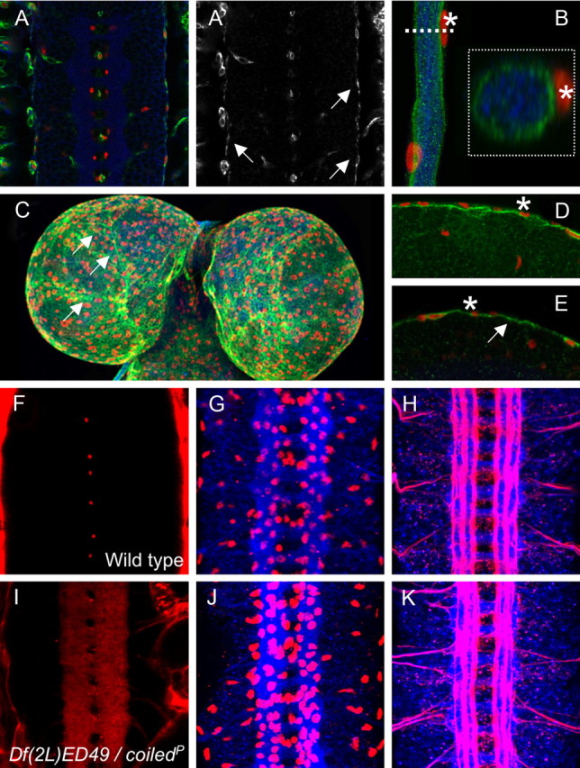
coiled does not affect overall CNS morphology. A, A′, Ventral nerve cord of a stage 16 CPTI001277 embryo carrying a GFP exon in the first intron of Coiled. GFP expression is shown in green, HRP expression to label all neuronal membranes is shown in blue, and glial nuclei are labeled by Repo staining in red. A′, GFP–Coiled expression. Coiled is expressed in the outer glial cells layer in the subperineurial glial cells (arrows). The GFP–Coiled protein accumulates in the cytoplasm and is not secreted into the extracellular space. B, Segmental nerve of a third-instar larva. GFP–Coiled is expressed by the subperineurial glia, which encircles the nerve. The nuclei abutting the GFP the subperineurial glia (asterisk) are nuclei of the perineurial glia. The boxed area is an orthogonal section taken at the level of the dashed line. C, Brain lobes of a third-instar larvae. Projection of a Z-stack. GFP expression is found in the subperineurial glia. Increased expression is noted at the cell boundaries (arrows). D, Single section showing expression in the subperineurial glia in the larval brain. The asterisks denote perineurial glia. E, Single section showing expression on the subperineurial glia in the adult brain (arrow). F, I, Frontal views on stage 17 nerve cords in live embryos. G, H, J, K, Frontal views on dissected stage 16 nerve cords stained for neuronal membranes (blue, anti-HRP staining), Repo expression (G, J, red), and Fasciclin2 expression (H, K, red). F, Wild-type stage 17 embryo injected with 10 kDa labeled dextran. No dye penetrates into the ventral nerve cord. G, H, Wild-type stage 16 embryos, showing normal pattern of glial cells and CNS axon tracts. I, Mutant coiled stage 17 embryo injected with 10 kDa labeled dextran. The dye penetrates into the nerve cord. J, K, Mutant coiled stage 16 embryos. Glial cell number and overall axon pattern are normal.
