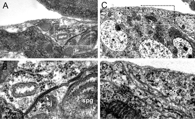
Figure 5.

Electron microscopic analysis of septate junction formation. Electron micrographs focusing on the CNS blood–brain barrier of late-stage 17 embryos. A, B, Wild-type embryo. The boxed area in A is shown in B. The subperineurial glial cells (spg) establish prominent electron-dense ladder-like septate junctions (sj, arrowhead). C, D, In coiled mutant embryos, no septate junctions can be detected.
