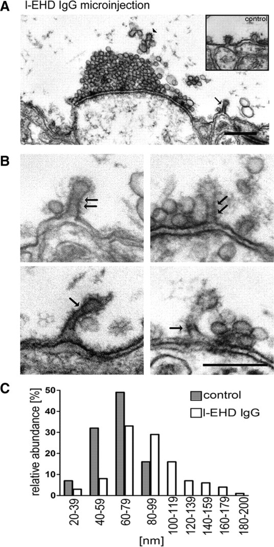
Figure 3.

Perturbation of l-EHD at synaptic release sites causes accumulation of coated pits with elongated necks. A, Electron micrograph of a synapse in a giant reticulospinal axon microinjected with l-EHD IgG followed by action potential stimulation at 5 Hz for 30 min. Note the clathrin-coated pit with an elongated neck (arrow; observed in 10 different antibody-injected giant axons in 3 different animals; coated pits in a control axons are shown in the inset). Clathrin coats visible on endosome-like structures above the synaptic vesicle cluster are indicated by an arrowhead. Scale bar, 0.5 μm. Axons microinjected with EHD antibodies, but maintained at rest in low-calcium Ringer, had a normal appearance (data not shown). B, Examples of coated pits in the periactive zone of giant reticulospinal axons injected with l-EHD IgG followed by action potential stimulation. Helix-like structures at the elongated necks are indicated with arrows. Scale bar, 0.2 μm. C, Distribution of the length of clathrin-coated pits in l-EHD IgG-injected axons (open bars, n = 117) and control axons (filled bars, n = 67).
