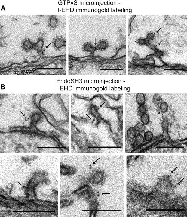
Figure 5.

Immunogold localization of l-EHD at coated endocytic structures trapped after perturbation of dynamin function. A, Examples of the localization of l-EHD on coated structures in the periactive zone of axons microinjected with GTPγS followed by action potential stimulation (Evergren et al., 2007). Gold particles are indicated by arrows. B, Examples of l-EHD localization on coated structures in the periactive zone of axons microinjected with the SH3 domain of endophilin followed by action potential stimulation (Gad et al., 2000). Scale bars, 0.15 μm.
