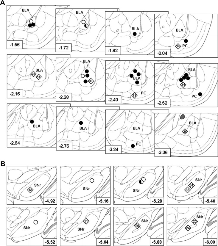
Figure 2.

Histologically verified kindling sites and single unit recording sites. A, Location of kindling electrodes in the right BLA or the piriform cortex (PC) in 30 rats, drawn on cutouts of coronal sections of the rat brain according to Paxinos and Watson (2007). Filled circles, Animals used for determination of the effect of VPA on kindled ADTs; rhombs, animals used for determination of the effect of VPA on kindled ADTs and for recording of the effect of VPA on SNr neurons (animal names are given within the rhombs to indicate animals that showed a poor or good response to VPA during electrophysiological recordings; compare with Fig. 4); half-filled circle, animal used for determination of the effect of VPA on kindled ADTs and for control recordings with injection of saline during electrophysiological recording; open circles, animals used for control recordings with injection of saline during electrophysiological recording. The distance to bregma in millimeters is given in the left corner of each section. B, Single-unit recording sites in the SNr in animals tested for the efficacy of VPA on SNr firing rates and patterns in kindled rats (rhombs; n = 7; animal names are given within the rhombs to indicate animals that showed a poor or good response to VPA during electrophysiological recordings; compare with Fig. 4) and saline in control rats (half-filled circle and open circles; together n = 4). The distance to bregma in millimeters is given in the right corner of each cutout of coronal sections of the rat brain according to Paxinos and Watson (2007).
