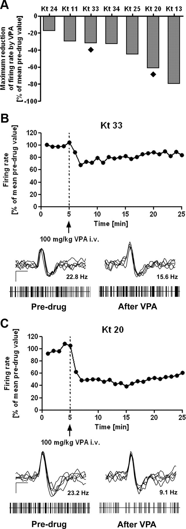
Figure 4.

Individual effect of valproate on spontaneous discharge rates. For evaluation of electrophysiological data, mean 1 min bins were calculated from firing rates relative to the mean individual predrug value (5 min of recording time before drug injection), which was set at 100%. Systemic application of VPA significantly reduced discharge rates of SNr neurons when all animals were considered (n = 7). Importantly, marked individual differences in response to VPA became evident. A, Comparison of maximum reduction of SNr discharge rates within 20 min after injection of VPA reveals marked interindividual differences between animals. The filled rhombs below bars indicate the two animals that were chosen to illustrate representative examples of VPA-induced effects on SNr firing in a poor (B) and a good (C) responder. Relative discharge rates are shown during 5 min before and 20 min after intravenous injection of 100 mg/kg VPA intravenous (dotted line). Representative recordings of the two sample neurons are also shown in B and C. Superimposed spikes (n = 5) of SNr neurons showing biphasic positive-negative waveforms as well as trains of discriminated spikes drawn as raster plots are shown for recordings before (predrug) and after (at the time of maximum reduction of firing rate) injection of VPA, respectively. Superimposed spikes (calibration: 0.5 ms, 25 μV) express biphasic positive-negative waveforms in predrug as well as in postdrug recordings. Raster plots (each with 5 s duration) reflect burst firing pattern independent of the presence of VPA in most of the recordings (refer to Table 4). The mean discharge rates of the shown neurons before or after injection of VPA are given above the trains.
