Figure 3.
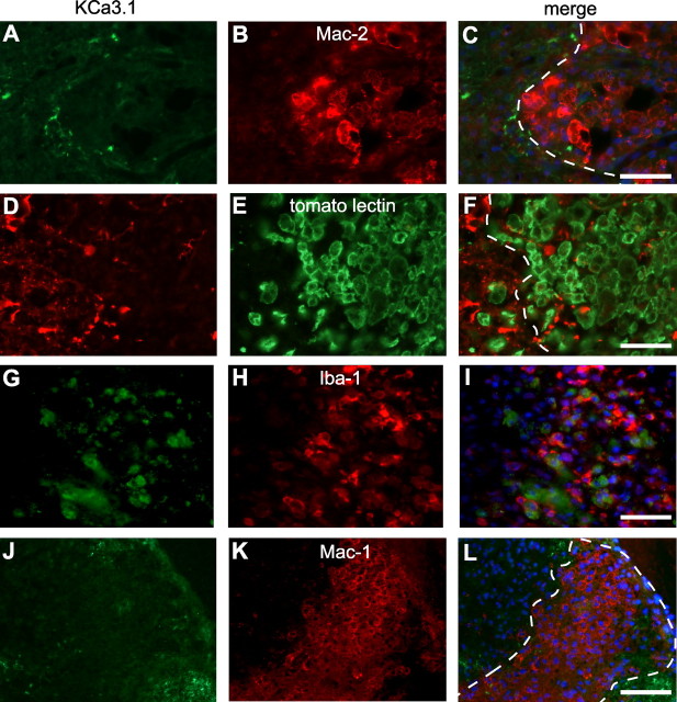
KCa3.1 immunoreactivity was not detected in macrophages and microglia in the injured mouse spinal cord. A–I, Double labeling with anti-KCa3.1 antibody (A, D, G) and different macrophage/microglial markers (B, E, H) in perfusion fixed tissue sections of the spinal cord at 7 d after SCI. The anti-Mac-2 antibody (B) recognizes phagocytic macrophages; tomato lectin (E) binds to macrophages and microglial cells; and Iba-1 (H) is upregulated in activated macrophages/microglia. The lesion core is to the right of the dashed lines in the merged images in C and F. Note that the macrophages/microglia in the lesion lack KCa3.1 staining. J–L, Unfixed tissue section of the spinal cord taken at 7 dpi was double labeled for KCa3.1 (J) and CD11b (Mac-1 antibody; K). Note the absence of double labeling of the Mac-1+ macrophages (L). Scale bars, 50 μm.

