
Figure 6.
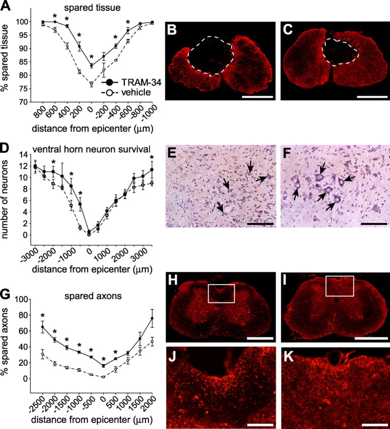
TRAM-34 treatment reduces secondary damage after SCI. All spinal cord histological sections were from mice at 28 dpi. A–C, Spinal cord sections taken at different distances from the lesion epicenter (at position 0) were labeled for GFAP, and the stained area was quantified (A). Data show significant sparing of tissue in TRAM-34-treated mice compared with vehicle-treated controls (p < 0.001). Micrographs show GFAP immunostaining at the epicenter of the injury in representative vehicle-treated (B) and TRAM-34-treated (C) mice. Note the smaller cystic cavity after treatment with TRAM-34. D–F, Tissue sections were stained with cresyl violet to quantify neurons in the ventral horn. Mice treated with TRAM-34 had greater neuron survival (D) in a region spanning 500-2000 μm rostral and 3000 μm caudal to the lesion epicenter (*p < 0.05). Micrographs showing cresyl violet staining of neurons in the ventral horn (arrows) at 500 μm rostral to the lesion epicenter in mice treated with vehicle (E) or TRAM-34 (F). G–K, Tissue sections were labeled for neurofilament 200 (NF200) to label axons. Mice treated with TRAM-34 had more spared axons (G) in the dorsal column white matter (*p < 0.001). Micrographs of NF-200 staining taken 500 μm rostral to the epicenter of the lesion. The areas outlined in the boxes for control (H) and TRAM-34-treated (I) mice are shown at higher magnification in J and K. More NF-200-stained profiles are present in the TRAM-34-treated mouse (K) than the vehicle-treated mouse (J). For all graphs, values are expressed as mean ± SEM (n = 6, TRAM-34 group; n = 8, vehicle group). Scale bars: B, C, H, I, 500 μm; E, F, J, K, 100 μm.