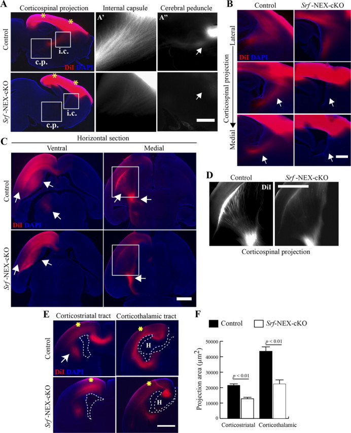
Figure 10.

Dil labeling shows impairment in axonal projections in Srf-NEX-cKO mutants. A, DiI crystals were placed on the brain surface in the regions of the motor and the visual cortices (indicated by asterisks) in P0.5 Srf-NEX-cKO knock-out and control littermates. Two weeks after labeling, brains were sectioned sagittally. Impaired corticospinal innervation was observed in the knock-out brain. Magnifications of the internal capsule (i.c) and cerebral peduncle (c.p) regions are shown in A' and A”. Projections through the cerebral peduncle are seen in the brains of control but Srf-NEX-cKO mice. B, Serial sagittal sections from lateral to medial regions of the brain show lack of corticostriatal projections (arrows) in Srf-NEX-cKO mice. No misguided axons were observed in the mutant mice. C, After 6 weeks of labeling, control and Srf-NEX-cKO brains were sectioned horizontally. Arrows show diminished projections within the neocortex, corticostriatal projections, and innervations to the thalamus in the mutant. Medial horizontal section shows impaired projections through the internal capsule and the cerebral peduncle. D, Magnified views of the boxed regions in C showing the corticospinal projections. E, Coronal sections from caudal regions of the brain reveal diminished corticostriatal as well as corticothalamic tracts (arrows). Asterisks indicate sites of crystal placement; dotted lines outline the ventricular zone and the hippocampus (H). F, Quantification of area of innervation by corticostriatal and corticothalamic axons in E (n = 3 mice).
