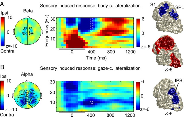
Figure 3.
Body-centered beta and gaze-centered alpha rhythms share a common substrate. A, Body-centered lateralization. Color format for all panels: warmer (red) colors, increase for targets to contralateral hand; cooler (blue) colors, increase for ipsilateral targets. Left, Scalp topography shows a beta power decrease (18–30 Hz) at central sensors (marked) during the sensory response period (t = 400 ms). Middle, Time-frequency resolved power changes for the marked sensors. Artifactual data indicated by a horizontal gray bar. Right, Top, Source reconstruction of the body-centered sensory response (18–30 Hz; t = 400 ms). Right, Bottom, Source reconstruction of the beta rebound during the delay period (t = 800 ms). B, Gaze-centered activity. Color format for all panels: warmer (red) colors, increase for targets in contralateral field; cooler (blue) colors, increase for ipsilateral targets. Left, Scalp topography shows an alpha power decrease (10 Hz; t = 400) at posterior and central sensors (marked). Middle, Time-frequency resolved power changes for the marked sensors. Right, Source reconstruction of the gaze-centered alpha-band suppression during the sensory response (10 Hz; t = 400 ms). Body-centered beta and gaze-centered alpha-band power share a common substrate in the SPL.

