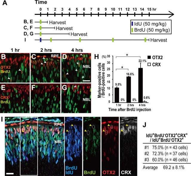
Figure 1.
OTX2 expression begins during the final RPC cell cycle. A, Schematic diagram of single or double S-phase labeling protocols used to administer thymidine analogs (IdU and/or BrdU) to label cells. Timed pregnant female mice at E14.5 were subjected to BrdU injection and at E15.5 were subjected to IdU and BrdU injection. B–G, Immunostaining of retinal section of embryos that were injected with BrdU 1 h (B, E), 2 h (C, F), or 4 h (D, G) before harvest. Retinal sections were stained with an anti-BrdU antibody and an anti-OTX2 antibody (B–D), or an anti-BrdU antibody and an anti-CRX antibody (E–G). The white arrowheads indicate BrdU-positive and OTX2-positive cells (B–D) or BrdU-positive and CRX-positive cells (G). RPE, Retinal pigment epithelium; NBL, neuroblastic layer. H, Ratio of the number of BrdU- and OTX2-positive cells (OTX2+/BrdU+), or BrdU- and CRX-positive cells (CRX+/BrdU+) to the total number of BrdU-positive cells, counted at each time point. Data are means ± SEM. n = 1505 cells from 9 sections (1 h, OTX2+/BrdU+), n = 524 cells from 3 sections (2 h, OTX2+/BrdU+), n = 519 cells from 4 sections (4 h, OTX2+/BrdU+), n = 1240 cells from 6 sections (1 h, CRX+/BrdU+), n = 1417 cells from 6 sections (2 h, CRX+/BrdU+), and n = 765 cells from 8 sections (4 h, CRX+/BrdU+). *p < 0.001, Student's t test. I, IdU- and BrdU-double-labeled retinas were immunostained with an anti-BrdU antibody, which stains both BrdU and IdU (blue, IdU- and/or BrdU-labeled cells); an anti-BrdU antibody, which stains only BrdU (green); an anti-OTX2 antibody (red); and an anti-CRX antibody (white). IdU was injected once 15 h before harvest while BrdU was injected every 4 or 5 h to label cells in S phase as represented in (A). The yellow arrowheads indicate IdU-positive, BrdU-negative, OTX2-positive, and CRX-positive cells delineated by the dotted circle. J, Percentage of IdU-positive, BrdU-negative, OTX2-positive, CRX-positive cells (IdU+/BrdU−/OTX2+/CRX+) in the IdU-positive, BrdU-negative, OTX2-positive (IdU+/BrdU−/OTX2+) cell population. Most IdU+/BrdU−/OTX2+ cells were CRX-positive, which is a postmitotic photoreceptor precursor marker. n = 126 cells on 11 sections of 4 retinas from 3 animals (#1, #2, and #3) were analyzed. Data are means ± SEM. Scale bars, 20 μm.

