
Figure 4.
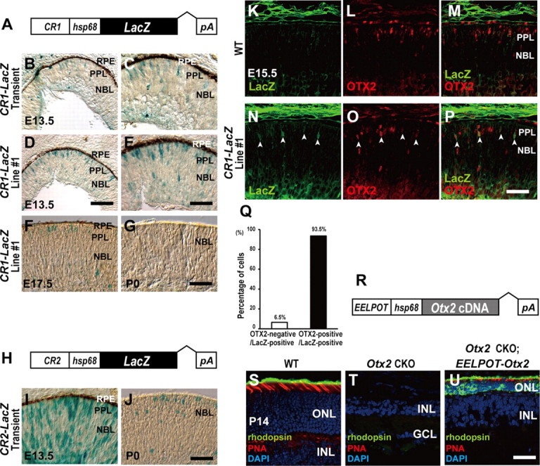
CR1 (EELPOT) recapitulates OTX2 expression in the embryonic mouse retina. A, Structure of CR1 (EELPOT)-LacZ reporter construct. B–G, X-gal staining of retinal sections from transient transgenic CR1 (EELPOT)-LacZ mice (B, C) and germline-transmitted CR1 (EELPOT)-LacZ transgenic mouse line (line 1) (D, E) at E13.5. (C, E), Higher magnifications of (B) and (D). X-gal staining of the retinal sections from E17.5 (F) or P0 (G) EELPOT-LacZ transgenic mouse line 1. RPE, Retinal pigment epithelium; PPL, presumptive photoreceptor layer; NBL, neuroblastic layer. Scale bars: D, 100 μm; E, G, 50 μm. H, Structure of CR2-LacZ reporter construct. I, J, X-gal staining of retinal sections from transient transgenic CR2-LacZ mice of E13.5 (I) or P0 (J). Scale bar: J, 50 μm. K–P, Immunostaining of retinal sections from WT (K–M) or line 1 mice (N–P). Immunostaining for LacZ (K, N), for OTX2 (L, O), and merge (M, P). The white arrowheads indicate both LacZ- and OTX2-expressing cells (N–P). Scale bar, 50 μm. Q, Percentage of OTX2-negative and OTX2-positive cells in LacZ-positive cells (n = 418 cells) in line 1 retinal sections at E15.5. R, Structure of EELPOT-Otx2 expression construct. S–U, Immunostaining of retinal sections from WT (S), Otx2 CKO (T), and transgenic mouse line, which expresses Otx2 under the control of EELPOT in the Otx2 CKO background (U). Sections were immunostained with an anti-RHODOPSIN antibody (green) and stained with PNA (red) and DAPI (blue) (S–U). Note that EELPOT-directed Otx2 expression in the Otx2 CKO retina resulted in a partial but significant rescue of Otx2 CKO phenotype (U). ONL, Outer nuclear layer; INL, inner nuclear layer; GCL, ganglion cell layer. Scale bar, 50 μm.