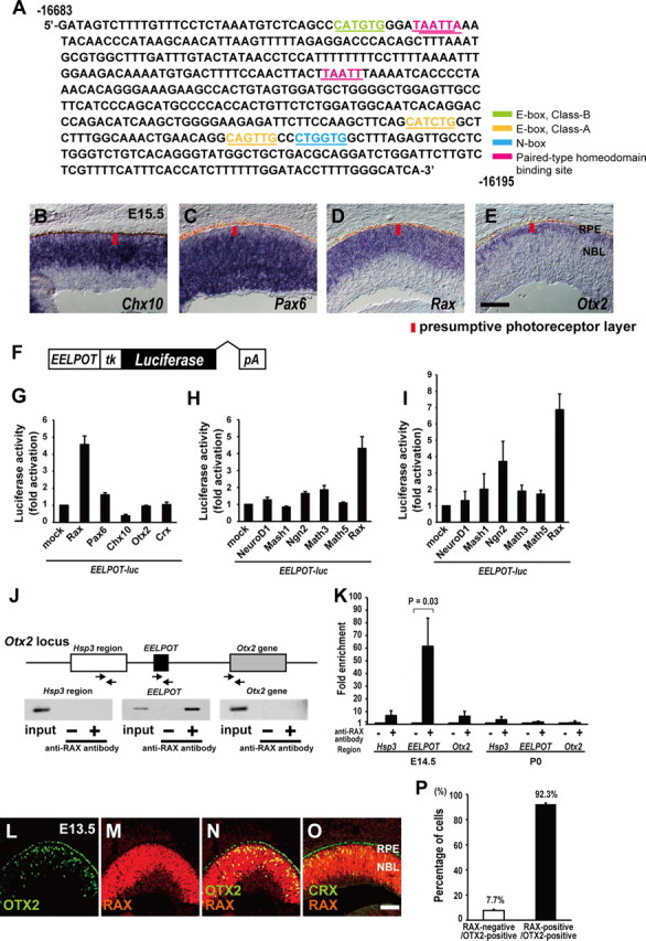
Figure 5.

RAX directly regulates Otx2 transcription in the embryonic mouse retina. A, DNA sequence of CR1 (EELPOT). Potential paired-type homeodomain-binding site (pink), E-box (class A) (orange), N-box (blue), E-box (class B) (green) are represented. B–E, Expression of Chx10 (B), Pax6 (C), Rax (D), and Otx2 (E) in the developing mouse retina at E15.5. RPE, Retinal pigment epithelium; NBL, neuroblastic layer. Scale bar, 100 μm. F, Structure of EELPOT-luc reporter construct. G–I, Luciferase reporter assay using EELPOT-luc. NIH3T3 cells (G, H) and Neuro2A (I) cells were transfected with EELPOT-luc reporter construct and expression plasmids of homeodomain transcription factors (G) or bHLH transcription factors (H, I). Data are means ± SEM (n = 3). J, Mouse Otx2 locus and primers for PCR of ChIP assay. The arrows represent primers for PCR. Mouse retinas at E15.5 were harvested and subjected to ChIP assay using the anti-RAX antibody. PCR was performed using primer set designed to 5′-upstream region of EELPOT (Hsp3 region), EELPOT, or Otx2 gene. K, Quantitative ChIP analysis both E14.5 and P0 for anti-RAX antibody. Data are means ± SEM (n = 4). Values of p by Student's t test. L–O, Immunostaining for OTX2 (L) and RAX (M). (N), Merge of (L) and (M). Coimmunostaining for RAX and CRX (O). Scale bar, 100 μm. P, Percentage of RAX-positive cells in OTX2-positive cells. n = 195 cells from 3 sections were analyzed. Data are means ± SEM.
