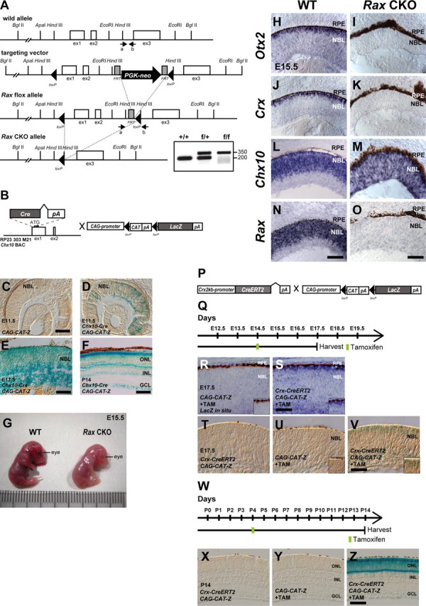
Figure 6.

Establishment and analysis of Rax flox, Crx 2 kb promoter-CreERT2, and Chx10-Cre mouse lines. A, Diagram of the Rax WT allele, the targeting vector, Rax flox allele, and Rax CKO allele. The white boxes, gray boxes, and arrowheads indicate exons, FRT sites, and loxP sites, respectively. PGK-neo indicates a phosphoglycerate kinase (PGK) promoter-driven neomycin-resistant gene expression cassette. The arrows indicate the positions of PCR primers used for genotyping. PCR products of 200 or 350 bp were amplified from WT or targeted allele, respectively. B, A schematic diagram of the modified BAC integrated with the Cre-pA cassette into the first exon of the mouse Chx10 gene, and CAG-promoter-directed LacZ construct. LacZ expression starts after the recombination occurs at loxP sites flanking chloramphenicol acetyltransferase (CAT)-pA cassette (CAG-CAT-Z). C–F, X-gal staining using sections from the retina of CAG-CAT-Z mouse at E11.5 (C) and from the retina of Chx10-Cre; CAG-CAT-Z mouse of E11.5 (D), E17.5 (E), or P14 (F). Scale bars: C, 200 μm; E, 50 μm; F, 100 μm. NBL, Neuroblastic layer; ONL, outer nuclear layer; INL, inner nuclear layer; GCL, ganglion cell layer. G, A picture showing mouse embryos at E15.5, WT (left) or Chx10-Cre; Raxflox/flox (Rax CKO) mouse (right). H–O, In situ hybridization of sections from the control (WT) retina (H, J, L, N) or Rax CKO retina (I, K, M, O) probed with antisense RNA of Otx2 (H, I), Crx (J, K), Chx10 (L, M), or Rax (N, O). Scale bars: H, J, L, N, 100 μm; I, K, M, O, 50 μm. P, Structure of Crx 2kb-promoter-driven CreERT2 construct and CAG-CAT-Z construct. Q, Schedule for tamoxifen injection and harvest of embryos. R–V, In situ hybridization probed with an antisense RNA against LacZ (R, S) or X-gal staining (T–V) of sections isolated from a Crx-CreERT2; CAG-CAT-Z mouse retina without tamoxifen (T) and CAG-CAT-Z mice (R, U) or Crx-CreERT2; CAG-CAT-Z mice (S, V) with tamoxifen. Tamoxifen was administrated to pregnant female mice at E14.5, and embryos were harvested at E17.5 as indicated in Q. The insets indicate the representative position of each panel. Scale bar, 50 μm. W–Z, X-gal staining using sections from a Crx-CreERT2; CAG-CAT-Z mouse retina without tamoxifen (X) and CAG-CAT-Z mice (Y) or Crx-CreERT2; CAG-CAT-Z mice (Z) with tamoxifen. Tamoxifen was administrated to mice at P4, harvested at P14 as indicated in (W), and subsequently processed for X-gal staining. Scale bar, 100 μm.
