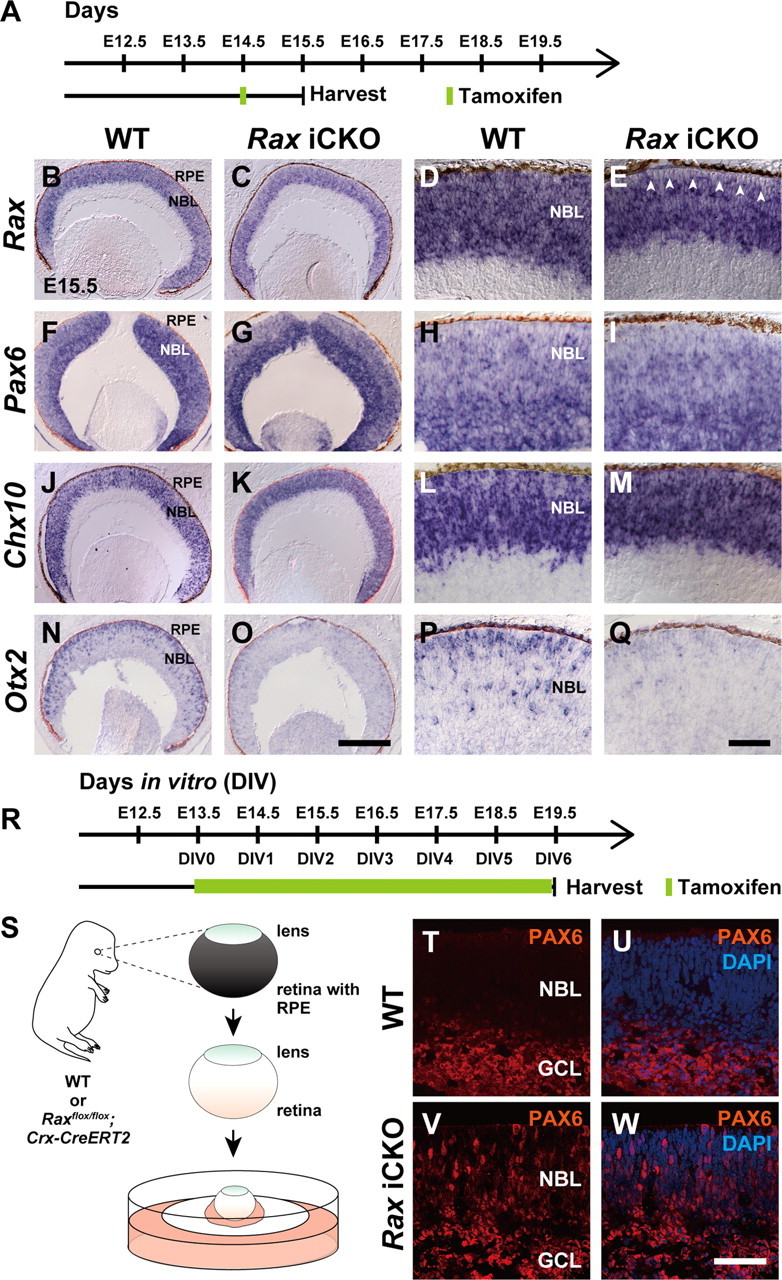
Figure 7.

Immunohistological analysis of the tamoxifen-induced Rax CKO (iCKO) retina. A, Schematic diagram of the schedule for tamoxifen injection and harvest of embryos. B–Q, In situ hybridization of sections from the control (WT) retina (B, D, F, H, J, L, N, P) or Rax iCKO retina (C, E, G, I, K, M, O, Q) probed with antisense RNA of Rax (B–E), Pax6 (F–I), Chx10 (J–M), and Otx2 (N–Q). D, E; H, I; L, M; and P, Q are higher magnifications of B, C; F, G; J, K; and N, O, respectively. The white arrowheads indicate the region where Rax gene was inactivated by tamoxifen-induced recombination (E). RPE, Retinal pigment epithelium; NBL, neuroblastic layer. Scale bars: O, 200 μm; Q, 50 μm. R, Schematic diagram of the schedule for tamoxifen treatment and harvest of explant cultures. S, E13.5 mouse retinas were used for explant culture after removal of RPE. Note that lenses were not removed. T–W, Immunostaining for PAX6 of the WT (T, U) or Rax iCKO (V, W) retina. Images of PAX6 immunostaining without (T, V) or with (U, W) DAPI counterstaining. GCL, Ganglion cell layer; NBL, neuroblastic layer. Scale bar, 50 μm.
