
Figure 2.
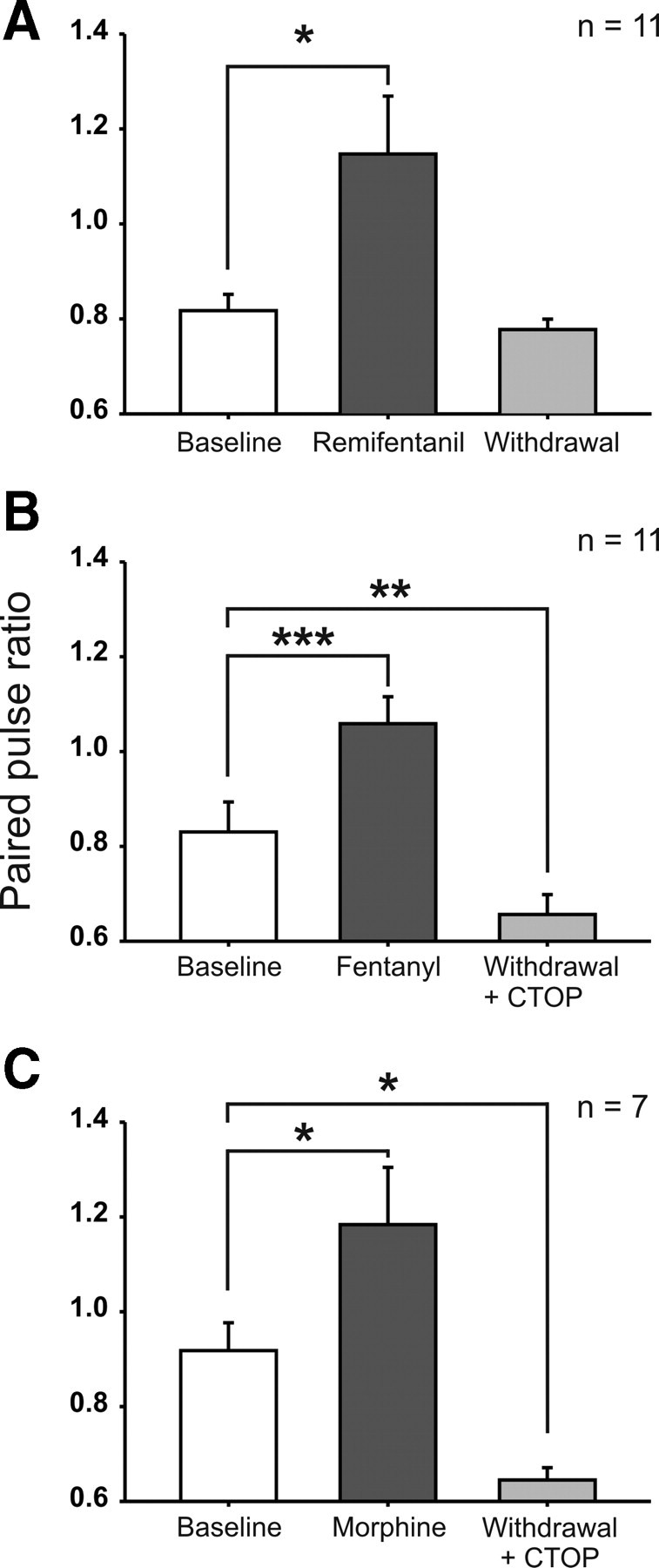
Intravenous opioids bidirectionally modulated the PPR of spinal C-fiber-evoked field potentials. Bar graphs represent the mean PPR during baseline (30 min before opioid infusion), during opioid infusion (at 15–45 min after onset of the infusion), and after withdrawal (at 15–45 min after termination of the infusion). A, During intravenous remifentanil infusion (dosing as in Fig. 1A), PPR was significantly increased and returned to baseline level after withdrawal. B, Fentanyl infusion (dosing as in Fig. 1B) was associated with an increased PPR. After withdrawal, precipitated by topic application of CTOP (10 μm), the PPR was depressed below baseline level. C, During morphine infusion (dosing as in Fig. 1C), the PPR increased significantly. After CTOP precipitated withdrawal, the PPR was decreased below baseline. *p < 0.05, **p < 0.01, ***p < 0.001, significant differences from baseline.