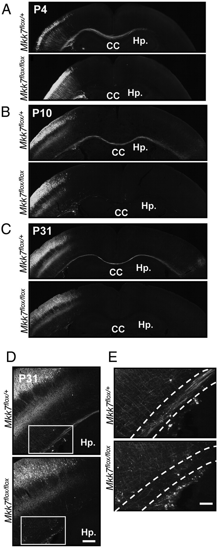
Figure 7.

Deletion of mkk7 in layer 2/3 neurons prevents axon elongation in vivo. A–C, Layer-specific MKK7 deletion. pCAG-NLS-Cre and pCAG-floxed-polyA-EGFP were introduced by in utero electroporation into the VZ of control Mkk7flox/+ and Mkk7flox/flox embryos at E15.5. Brains were fixed on the indicated postnatal days and coronal sections of the cortex were immunostained with anti-GFP antibody. Hp., Hippocampus. Scale bar, 1 mm. D, Higher-magnification view of axons in the cortices of the Mkk7flox/+ and Mkk7flox/flox mice in C. Scale bar, 200 μm. E, Higher-magnification view of the inset boxes in D, focusing on the white matter (broken lines). Scale bar, 100 μm.
