Figure 4.

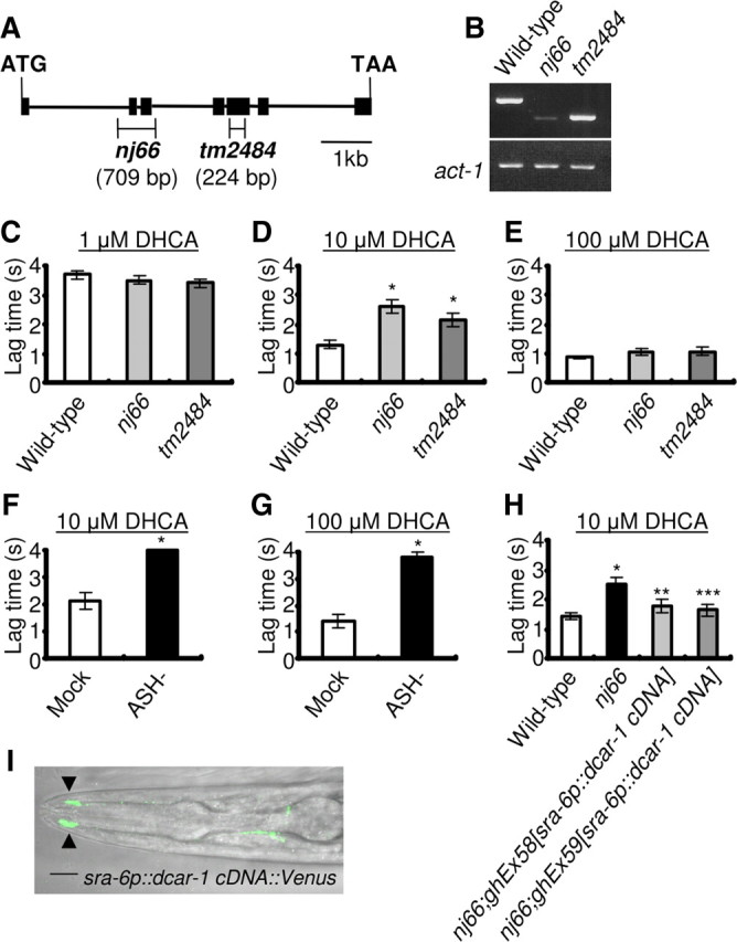
dcar-1 mutant animals show a defect in avoidance of DHCA. A, Structure of the dcar-1 gene. The locations and extent of dcar-1(nj66) and dcar-1(tm2484) deletions are indicated. Black boxes indicate exons; lines indicate introns. B, RT-PCR of total wild-type and dcar-1 mutant animals mRNA. act-1 messages were amplified in parallel as a positive control. C–E, Avoidance assays for DHCA. A small drop of DHCA buffer was placed in the path of an animal as it moved forward on agar plate. Average lag time for an animal to reverse direction after contacting with the DHCA was measured. If animals did not reverse within 4 s, the lag time was scored as 4 s. Data are shown as mean ± SEM (1 μm DHCA: wild-type, n = 31; nj66, n = 31; tm2484, n = 28; 10 μm DHCA: wild-type, n = 34; nj66, n = 35; tm2484, n = 32; 100 μm DHCA: wild-type, n = 33; nj66, n = 34; tm2484, n = 33). Asterisks indicate significant differences from wild-type animals (*p < 0.01 by one-way ANOVA). F, G, ASH-ablated animals showed a defect in the 10 and 100 μm DHCA avoidance. Data are shown as mean ± SEM (10 μm DHCA: mock-ablated, n = 9; ASH-ablated, n = 10; 100 μm DHCA: mock-ablated, n = 10; ASH-ablated, n = 11). Asterisks indicate significant differences from mock-ablated animals (*p < 0.001 by Student's t test). H, The 10 μm DHCA avoidance defect of dcar-1(nj66) mutant animals was rescued by the expression of dcar-1 cDNA in a subset of neurons including ASH neurons using the sra-6 promoter in two independent transgenic animals. Data are shown as mean ± SEM (wild-type, n = 46; nj66, n = 48; nj66;ghEx58[sra-6p::dcar-1 cDNA], n = 22; nj66;ghEx59[sra-6p::dcar-1 cDNA], n = 24). Asterisks indicate significant differences from wild-type animals (*p < 0.01 by one-way ANOVA) and dcar-1(nj66) mutant animals (**p < 0.05, ***p < 0.01 by one-way ANOVA). I, Confocal image of sra-6p::dcar-1::Venus expression in the head of larva. Black arrowheads indicate the sensory cilia of the head. Scale bar, 10 μm.
