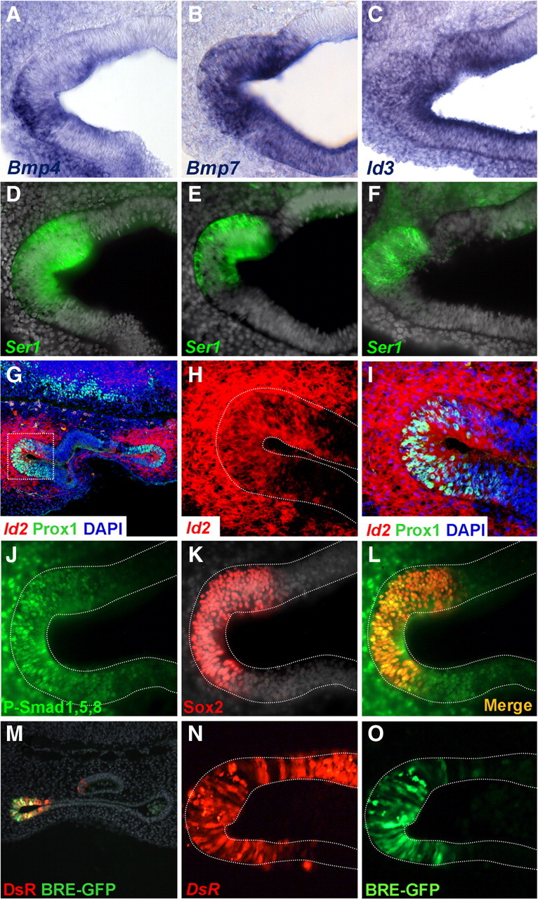
Figure 1.

Bmp pathway and Id gene expression in the prosensory patches. Coronal sections of E4 embryos show prosensory domains of vestibular cristae. A–F, Bmp4, Bmp7, and Id3 expression in the prosensory patches. Otocysts were processed for ISH with Bmp4 (A), Bmp7 (B), or Id3 (C) riboprobes and for Serrate1 immunofluorescence (D–F). G–I, Id2 gene expression in the prosensory patches. Confocal images of coronal sections processed for ISH with Id2 probe (red) and for immunofluorescence for Prox1 (green). The in situ hybridization signal was captured by confocal microscopy in the near infrared, Id2 was expressed in two epithelial regions corresponding to the prosensory domains of the cristae and the surrounding mesenchyme (G). An enlargement of the boxed area is shown in H and I to better illustrate the superposition of Id2 and Prox1. J–L, Immunofluorescence for P-Smad1,5,8 (J) and Sox2 (K) counterstained with DAPI. The merged image is shown in L. Note that P-Smad1,5,8 immunoreactivity overlaps the prosensory patch as identified by Sox2 expression, but it also extends outside the Sox2 domain into the neighboring epithelium and the surrounding mesenchyme. M–O, Smad transcriptional activity in prosensory patches. Cryostat section of the otic vesicle with the DsRed and the EGFP signal is shown in M. The prosensory domain of the anterior crista exhibited DsRed (N) and BRE directed EGFP signals (O). Note that the extension of the DsRed signal is broader than the BRE directed EGFP signal that is restricted to the anterior pole of the otic vesicle. For better comparison, photographs from different preparations were flipped horizontally to show either the anterior or posterior cristae with the same orientation: A–F, M–O, anterior cristae; G–L, posterior cristae; medial is always to the top.
