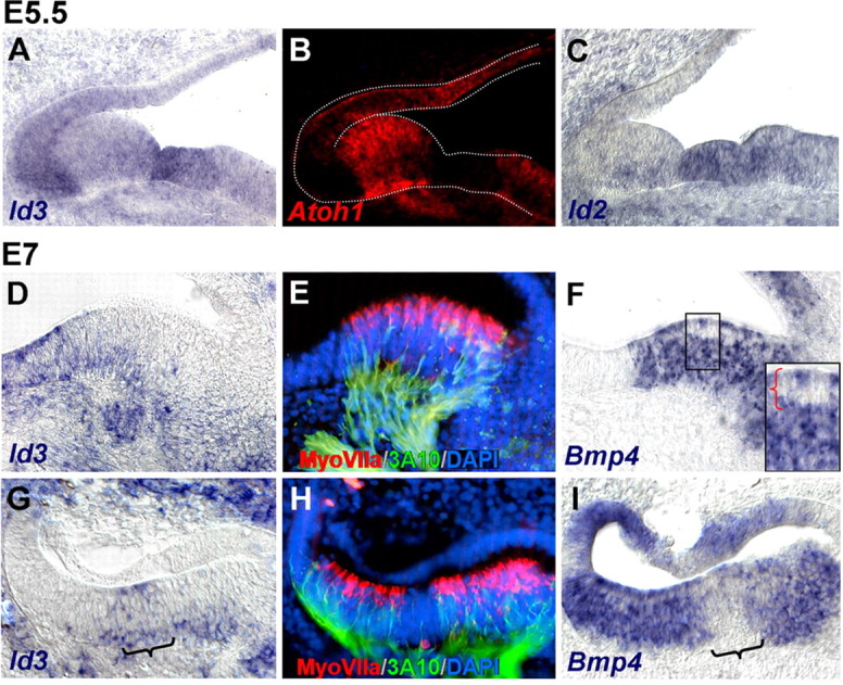
Figure 4.

Bmp and Id expression during differentiation of the sensory patches. A–C, Transversal alternate sections of the inner ear of E5.5 embryos, at the level of the lateral crista, double-labeled for Id3 (A) and Atoh1 (B) by fluorescent double ISH, and for Id2 (C). Atoh1 and Id3 were expressed complementary to each other. D–I, Transversal alternate sections of the inner ear of E7 embryos, at the level of the lateral (D–F) and posterior (G–I) cristae, probed for Id3 (D, G) and Bmp4 (F, I) by ISH. The brackets in G and I indicate the expression of Id3 in nonsensory epithelium devoid of Bmp4 signal, the cruciatum. The box in F shows a detail of a region containing a row of hair cells (red bracket), with reduced or absent Bmp4 signal. E and H are the same as D and G, which were colabeled with MyoVIIa (red) and 3A10 (green) antibodies to better identify the sensory domain and the developing hair cells. Nuclei were counterstained with DAPI (blue).
