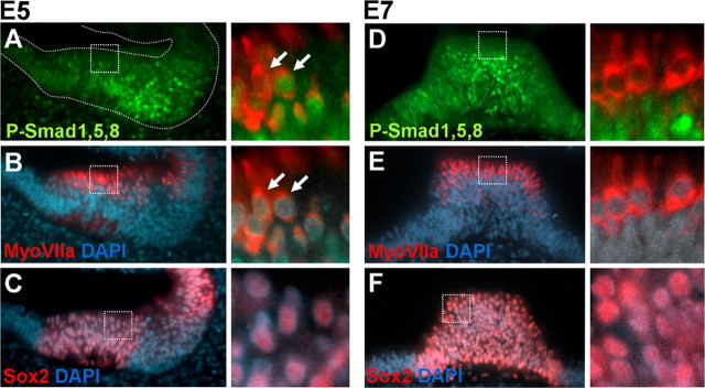
Figure 5.
P-Smad1,5,8 in sensory organs. A–C, Coronal section of E5 otocysts showing the anterior crista colabeled for P-Smad1,5,8 (A, green) and MyoVIIa (B, red). The corresponding alternate serial section was stained for Sox2 (C, red). Sections were counterstained with DAPI (blue). The details show the double-labeling with MyoVIIa and P-Smad1,5,8 and MyoVIIa and DAPI. Note that at this stage, P-Smad1,5,8, was downregulated from some but not all hair cells. The detail of Sox2 expression in the patch is shown for comparison (detail of C). D–F, Coronal section of E7 otocysts showing the posterior crista colabeled for P-Smad1,5,8 (D, green), MyoVIIa (E, red), and DAPI and corresponding alternate section stained for Sox2 (F, red). A detail of the double-labeling with MyoVIIa and P-Smad1,5,8 and MyoVIIa and DAPI are shown to the right. At this stage, hair cells did not express P-Smad1,5,8.

