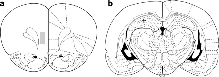
Figure 1.
a, mPFC electrodes targeted deep layers of prelimbic and infralimbic cortices (+3.2 mm AP and +0.6 mm ML to bregma; 3.2 mm DV from dura). The shaded box represents the region within which all the electrode tracts were located based on histology, starting coordinates, and distance that electrodes were moved. The cross in b shows the location of a typical lesion site marking the tip of the single EEG electrode in the dorsal hippocampus (CA1) (−3.8 mm AP and +2.5 mm ML to bregma; 2.5 mm DV from dura).

